ISSN : 0975-3710
EISSN : 0975-9107

JAYALAKSHMI N.R.1*, SARASWATHI K.J.T.2, VIJAYA B.3, RAMAN D.N.S.4, SHREENIVAS D.P.H.S.5
1Department of Microbiology and Biotechnology, Jnana Bharathi Campus, Bangalore University, Bangalore-56
2Department of Microbiology and Biotechnology, Jnana Bharathi Campus, Bangalore University, Bangalore-56
3Department of Microbiology and Biotechnology, Jnana Bharathi Campus, Bangalore University, Bangalore-56
4Department of Botany, P.G. Centre, St. Josephs College, Bangalore-12
5Lotus Labs Pvt Ltd., Bangalore-51
* Corresponding Author : jb_urs@rediffmail.com
Received : 26-07-2011 Accepted : 16-08-2011 Published : 30-09-2011
Volume : 3 Issue : 2 Pages : 97 - 102
Int J Agr Sci 3.2 (2011):97-102
DOI : http://dx.doi.org/10.9735/0975-3710.3.2.97-102
Conflict of Interest : None declared
Acknowledgements/Funding : I am greatly indebted to Prof. B.B. Kaliwal, Chairman, P. G. Department of Studies in Biotechnology and Microbiology, Karnatak University, Dharwad-580 003, for his review and correction of this paper and I am also grateful to Suresh. R, Department of Stat
The changes of growth pattern and anthocyanin production, malvidin and delphinidin of Malva sylvestris L., were studied under UV-B treatment. The supplemental dose of UV-B exposure significantly reduced the growth of the plant with increase in anthocyanin content. With the traditional chilled acidified methanol method, the total anthocyanins were extracted and estimated by pH differential spectroscopic method. The anthocyanins were purified by C18 Sep-pak column and further analyzed for different anthocyanins by high performance liquid chromatography (HPLC) methods.
Phenolics, malvidin, delphinidin, photoinduction, morphological variations, induced stress, HPLC, C18 Sep-pak column.
The interaction between UV-B radiation and plants are various like additive, synergistic or antagonistic [1] . Different species have different response to the level of UV-B radiation. The negative effects results in deformed morphological parameters like decrease in plant height, leaf area, leaf number, wet and dry mass of the plant, axillary branching etc., with increase in leaf curling. The positive effect is shown by enhanced acceleration in the biosynthesis of phenolics [2] .
Phenolicsin the epidermal layers of leaves and tissues are susceptible to UV light and protect the plants from UV damage by activating the enzymes chalcone synthase (CHS) or chalconeisomerase (CHI) in flavonoid biosynthetic pathway. This photoinduction activates photoreceptors, phytochromes and independently induces the synthesis of anthocyanins [3] .
Malva sylvestris L., (mallow) is a perennial herb of Malvaceae family harbors polysaccharides, flavonoids with anthocyanins as the main components. The alcoholic extracts of leaves and flowers have been widely used as a mild relief for cough and inflammatory diseases of the mucus membrane [4] , they are also utilized as medicines, food flavors, nutritive food, UV protecting agents (lotions and creams) etc. in pharma industries [5] . The prime aim of the present work was to study the effect of UV-B radiation on the growth and higher accumulation of anthocyanin production in Malva sylvestris.
Malvidin-3-glucoside and Delphinidin chloride were purchased from Sigma Aldrich (Germany), Solid phase extraction columns C18 Sep-pak columns from Agilent (USA), and all other HPLC graded chemicals from Himedia (India).
Malva sylvestris plants were cultivatedfrom seeds sown using top soil with mixture of compost to maintain moisture at room temperature. The flowering plants (ten numbers) were taken for elicitation. The UV-B radiation was provided for thirty minutes in three days by Philips sunlamps (Philips TL 20W/12). The sunlamps were wrapped by 0.13mm cellulose diacetate filters for removing UV-C radiation. The plants were observed for their morphological changes and later the treated flowers were dried under shade and stored at 4oC till further analysis.
Different methods were adopted for maximum recovery of anthocyanins. As per the percentage of yield, extraction and purification were done by the method as explained by [6] . Methanol:Aceticacid:Water in the ratio 49:1:50 was added to the powdered flower sample and incubated at 40C for 20-24 hours. The extract was filtered with Whatmann no.1 filter paper and the residual extracts were rotary evaporated under vacuum at 300C. The anthocyanins were separated by solid phase extraction using Accu Bond C-18 cartridge (sep-pak column) with acidified (0.1%Hcl) methanol as a solvent.
The total monomeric anthocyanin content was determined by pH differential spectrophotometric analysis with cyanidin as standard [7] and the purified sample with standard malvidin and delphinidin were qualitatively separated by TLC on silica gel 60 F254 with the solvent system butanol; aceticacid;water in the ratio 4:1:5. The pink and blue colored bands were obtained and their Rf values were calculated and compared with the standard.
Reverse phase HPLC analysis was carried out on waters separation module (Waters Corp., Milford, Mass., USA) equipped with an auto injector and separation was carried on ODS column. The sample was eluted at a flow rate of 1.5ml/min with two solvent system, solvent A with 15% acetic acid and 85% water (v/v), solvent B with acetonitrile. Gradient separation at room temperature was done with detection at 520nm.
Calculations and statistical analysis were performed using SPSS 11.5 Windows software. Based on the experimental design adopted in the study, data were analyzed using Student’s t-test. The results presented are averaged over the independent experiments with ten quantifications within each sample. Mean values are expressed as ± S.E. for comparison of the means, the at 1% level of significance.
The treatment given for three days, thirty minutes was non lethal for plant survival with flowering [Table-1] .
The UV treated plants showed morphological changes inplant height; leaf texture, number, area; fresh and dry weight of the plant; size and color of flowers. About 74% and 90% reduction in fresh and dry weight biomass, 63% reduction in plant height, 85% reduction in leaf number, 88% reduction in leaf area were noticed. The leaf texture showed roughness with curling and flower showed deepened blue color with size reduction [Table-2] , [Fig-1] and [Fig-2] .
The reduction in plant height is often used to index the degree of UV-B sensitivity, the basis is the free radicals being synthesized due to stress which change the membrane integrity; disrupts the synthesis and transport of plant hormones. The reduction in fresh and dry weights could be due to deficiency in photosynthetic activity with decreased efficiency of enzyme activities and photosystem II [8] . According to [9] the reduction in leaf area with decrease in leaf number could be an adaptive mechanism to minimize the exposure to radiation. The reduction can also be due to inhibited or reduced cell division, photosynthetic rate and calmodulin content, a key factor for leaf growth [10] . This similar trend in morphometric variations have also been observed in [11] Jaborosa magellanica [12] , Crotalaria juncea [13] , Oleaeuropaea [14] and Gossypium hirsutumL. [15] .
The solvent extraction of anthocyanin is the initial step to determine the total and individual anthocyanins present prior to quantification, purification, separation, and characterization [16] . In the present study extraction of anthocyanins was maximum (95%) with 1% acidified methanol by cold temperature incubation method when compared to Soxhlet (60-70%) and partition extraction (50-60%) methods [Table-3] .
The polar character of anthocyanin makes them soluble in several types of solvents such as methanol, ethanol, acetone, water etc., and generally involves the use of acidified methanol or ethanol. The use of acid stabilizes anthocyanins in the flavyliumcation form, which is red at low pH. Even though methanol is toxic, it is best preferred for complete extraction because ethanol is less efficient and more difficult to eliminate later during purification [17,18] .
The total anthocyanin concentration was significantly increased to about 180.08% compared to untreated sample [Table-2] and [Fig-2] . This increase justifies the work done in other plants like Arabidopsis, Lettuce, Petunia, Vitis vinifera, Catharanthus roseus, Passiflora quadrangularis,Crotalaria juncea L. etc. [3,19-23,13] . According to [24] flavonoid accumulation is regarded as a protective mechanism in higher plants. UV-B radiation induces injury to plant cells and is associated with alteration of oxidative stress defensive systems which result in disturbance of active oxygen metabolism with increase in scavengers like glutathione and possibly flavonoids, especially the anthocyanins [10] . Anthocyanins are water soluble pigments appearing transiently in juvenile or senescing tissues [7] and regulate the stress by reducing UV-B penetration, protect photosynthetic apparatus, bind to phytotoxins, and reorients cell division apparatus [25,26] .
Fractionation of acidified methanolic extract of flowers by Sep-pak C18 column afforded two anthocyanidins viz., malvidin and delphinidin which were identified by TLC and HPLC methods. The HPLC chromatogram of the dried flower sample extract obtained in the visible spectral region (520nm) revealed the presence of malvidin and delphinidin in both UV treated and untreated plants [Fig-3] and [Fig-4] .
The absorption spectra of the solutions showed the maxima to be identical (517nm), confirming the presence of the same anthocyanidins with the standard. According to the area of the corresponding peaks there was an increase in malvidin and delphinidin content in treated samples (49.3µg/g) and (27.4µg/g) when compared to untreated samples (27.9µg/g) and (14.2µg/g). This justifies the work done by [5] , which reveals Malva sylvestris is rich in malvidin and its derivatives compared to delphinidin.
The methanolic extracts of Malva sylvestris has traditionally been used as herbal medicine in folk remedies for the treatment of coughs, pains, inflammations and cancers. The plant could be used for the extraction of anthocyanins intended to be employed as food colorant and antioxidant agent by the food, pharmaceutical, and cosmetic industries.
The procedures adopted in this study can be used prior to and in conjugation with acid hydrolysis to fully characterize the malvidin and delphinidin derivitives. For complete identification of these compounds additional techniques like, electroscopy mass spectroscopy, NMR are necessary for determining the molecular weight, nature of glycosidic linkages and the sites of acyl and sugar substitution.
I am greatly indebted to Prof. B.B. Kaliwal, Chairman, P. G. Department of Studies in Biotechnology and Microbiology, Karnatak University, Dharwad-580 003, for his review and correction of this paper and I am also grateful to Suresh. R, Department of Statistics, Bangalore University for his expertise assistance in applying statistical tools for the present study.
[1] Petropoulou Y., Kyparissis A., Nikolopoulos D. and Manetas Y. (1995)Physiol. Plant, 94: 37–44.
» CrossRef » Google Scholar » PubMed » DOAJ » CAS » Scopus
[2] Zuk-Golaszewska K., Upadhyaya M.K., Golaszewski J. (2003) Plant Soil Environ, 49(3): 135–140.
» CrossRef » Google Scholar » PubMed » DOAJ » CAS » Scopus
[3] Li J., Ou-Lee T.M., Raba R., Amundson R.G. and Last R.L. (1993) Plant Cell, 5:171–179.
» CrossRef » Google Scholar » PubMed » DOAJ » CAS » Scopus
[4] Farina A., Doldo A., Cotochini V., Rajevic M., Quaglia M., Mulinacci N. and Vincieri F.F. (1995) J Pharm Biomed Anal, 14:203-211.
» CrossRef » Google Scholar » PubMed » DOAJ » CAS » Scopus
[5] Anderson O.M. and Markham K.R. (2006) CRC Press, Boca Raton, pp 471−551.
» CrossRef » Google Scholar » PubMed » DOAJ » CAS » Scopus
[6] Guisti M., Rodriguez S.L., Griffin D. and Wrosland R. (1999) J. Agric. Food Chem, 47: 4657-4664.
» CrossRef » Google Scholar » PubMed » DOAJ » CAS » Scopus
[7] Mazza G., Cacace J.E. and Kay C.D. (2004) J AOAC Int, 87: 129-145.
» CrossRef » Google Scholar » PubMed » DOAJ » CAS » Scopus
[8] Biggs R.H., SissonW.B. and Caldwell M.M. (1975) Nat. Techn. Info. Serv. Springfield, VA., pp. 263-273.
» CrossRef » Google Scholar » PubMed » DOAJ » CAS » Scopus
[9] Krizek D.T., Mirecki R.M. and Kramer G.F. (1994) Physiol Plant, 90: 593–599.
» CrossRef » Google Scholar » PubMed » DOAJ » CAS » Scopus
[10] Dai Q., Yan B., Huang S., Lui X., Peng S., Miranda L.L., Chavez A.Q., Vergara B.S. and Olszyk D.M. (1997) Physiol Plant, 101:301–308.
» CrossRef » Google Scholar » PubMed » DOAJ » CAS » Scopus
[11] Singh A. (1996) C. J. Bot., 74: 135-139.
» CrossRef » Google Scholar » PubMed » DOAJ » CAS » Scopus
[12] Cuadra P., Herrera R. and Fajardo V. (2004) Journal of Photochemistry and Photobiology, 76: 61–68.
» CrossRef » Google Scholar » PubMed » DOAJ » CAS » Scopus
[13] Balakrishnan V., Ravindran K.C., Venkatesan K. and Karuppusamy S. (2005) EJEAFChe, 4 (6):1125-1131.
» CrossRef » Google Scholar » PubMed » DOAJ » CAS » Scopus
[14] Liakoura V., Stavrianakou S., Liakopoulos G., Karabourniotis G. and Manetas Y. (1999) Tree Physiology, 19: 905—908.
» CrossRef » Google Scholar » PubMed » DOAJ » CAS » Scopus
[15] Zhao D., Reddy K.R., Kakani V.G., Read J. andSullivan J.H. (2003) Plant cell and environment, 26(5): 771-782.
» CrossRef » Google Scholar » PubMed » DOAJ » CAS » Scopus
[16] Durst R.W. and Wrolstad R. (2001) New York, Wiley, pp. F1.3.1–F1.3.13.
» CrossRef » Google Scholar » PubMed » DOAJ » CAS » Scopus
[17] Feluki T. (1969) J. Food Sci, 34: 365-349.
» CrossRef » Google Scholar » PubMed » DOAJ » CAS » Scopus
[18] Rivas-Gonzalo J.C. (2003) Cambridge: The royal society of Chemistrypp. 338– 358.
» CrossRef » Google Scholar » PubMed » DOAJ » CAS » Scopus
[19] Ong W.D. and Chong K.P. (2009) Modern Applied Science, Canadapp. 124-234.
» CrossRef » Google Scholar » PubMed » DOAJ » CAS » Scopus
[20] Ryan K.G., Swimmy E.E., Winefield C. and Markham K.R. (2001) Z Naturforsh, 56c: 745-754.
» CrossRef » Google Scholar » PubMed » DOAJ » CAS » Scopus
[21] Zhang W., Curtin C., Kikuchi M. and Franco C. (2002) Plant Sci, 162: 459-468.
» CrossRef » Google Scholar » PubMed » DOAJ » CAS » Scopus
[22] Vazquez-Flota F.A. and De Luca V. (1998) Phytochem, 49: 395-402.
» CrossRef » Google Scholar » PubMed » DOAJ » CAS » Scopus
[23] Antognoni F., Zheng S., Pagnucco C., Baraldi R., Poli F. and Biondi S. (2007) Fitoterapia, 78:345-352.
» CrossRef » Google Scholar » PubMed » DOAJ » CAS » Scopus
[24] Tevini M., Iwanzik W. and Thoma U. (1981) Planta, 153: 388–394.
» CrossRef » Google Scholar » PubMed » DOAJ » CAS » Scopus
[25] Pal M., Sengupta U.K., Srivastava A.C., Jain V. and Meena R.C. (1999) Plant Physiol, 4: 79-84.
» CrossRef » Google Scholar » PubMed » DOAJ » CAS » Scopus
[26] Winkel-Sherley B. (2002) CurrOpin Plant Biol, 5: 218-223.
» CrossRef » Google Scholar » PubMed » DOAJ » CAS » Scopus
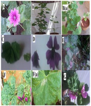 | Fig. 1- Effect of UV on Malvasylvestris A: Normal plant, B and Ba: Comparison of untreated and treated plant, C: Comparison of untreated and treated leaves, D: Comparison of untreated and treated flowers, E: Flowers with deep blue color, F and Fa: Leaf curling, G: UV treated plant with changes in morphology |
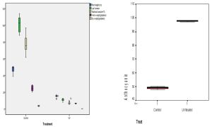 | Fig. 2- a and b. Statistical box plots showing the effect of UV treatment on physical characters and anthocyanin content The p-value is less than 0.01, the hypothesis is rejected at 1% level of significance and hence there is a significant difference between the treated and control plant |
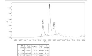 | Fig. 3- Chromatogram for untreated sampleand UV treated sample |
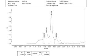 | Fig. 4- Chromatogram for untreated sampleand UV treated sample |
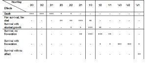 | Table 1- Survivability of Malva sylvestris with UV-B treatment +++: All ten plants, ++: Half of the plants, +: One or two plants |
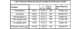 | Table 2- Effect of UV radiation on plant morphology and Anthocyanin content At 1% level of significance |
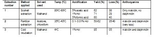 | Table 3- Recovery of total anthocyanin (by different extraction methods and conditions) |