ISSN : 0975-3710
EISSN : 0975-9107

MARTINS J.L.1*, XAVIER D.M.2, SILVA A.S.3, SANTOS R.P.4, MESKO M.F.5, COSTA S.N.6, FREIRE V.N.7, CAVADA B.S.8
1Departamento de QuÃmica AnalÃtica e Inorgânica, Universidade Federal de Pelotas, Pelotas, Rio Grande do Sul, Brazil.
2Departamento de QuÃmica AnalÃtica e Inorgânica, Universidade Federal de Pelotas, Pelotas, Rio Grande do Sul, Brazil.
3Departamento de QuÃmica AnalÃtica e Inorgânica, Universidade Federal de Pelotas, Pelotas, Rio Grande do Sul, Brazil.
4Laboratório de Engenharia de Materiais e da Computação de Sobral (LEMCS), Universidade Federal do Ceará, Sobral, Ceará, Brazil.
5Departamento de QuÃmica AnalÃtica e Inorgânica, Universidade Federal de Pelotas, Pelotas, Rio Grande do Sul, Brazil.
6Departamento de FÃsica, Universidade Federal do Ceará, Fortaleza, Ceará, Brazil.
7Departamento de FÃsica, Universidade Federal do Ceará, Fortaleza, Ceará, Brazil.
8Laboratório de Moléculas Biologicamente Ativas, Departamento de BioquÃmica, Universidade Federal do Ceará, Fortaleza, Ceará, Brazil.
* Corresponding Author : jmartins.martins@gmail.com
Received : - Accepted : - Published : 25-06-2012
Volume : 4 Issue : 5 Pages : 238 - 242
Int J Agr Sci 4.5 (2012):238-242
DOI : http://dx.doi.org/10.9735/0975-3710.4.5.238-242
Conflict of Interest : None declared
Humic acid (HA) is a principal component of humic substances, which are the major organic constituents of soil, peat, coal, many upland streams, dystrophic lakes and ocean water. HA has an important impact on a variety of environment processes. This work aimed to characterize the HA extracted and purified from the Candiota coalfield (Brazil) by visible (VIS), Fourier transform infrared (FTIR), energy dispersive X-ray (EDX), inductively coupled plasma absorption emission (ICP-AES), and X-ray diffraction (XRD) spectroscopies, thermogravimetry (TG), differential thermal analysis (DTA), and scanning electron microscopy (SEM). The results of these analyses showed that Candiota HA presents a high degree of humification, which is considered to be type A with a turbostratic structure. The elemental analysis (EDX/ICP-AES) indicated the presence of low concentrations of heavy metals.
Humic acid, coal, characterization, spectroscopy, thermal analyses.
The organic matter of soils can be divided into non-humic and humic substances. Carbohydrates, amino acids, protein, lipids, nucleic acids, and lignins are non-humic substances that originate from plants and other organisms. However, humic substances (HSs) are materials synthesized during the decomposition of plants and animal residues with or without the assistance of microorganisms (humification). HSs are defined as hydrophilic, acidic, high molecular weight, amorphous, and colloidal polydispersed substances with a yellow to brown-black color [1] HSs are not only found in soils, but are also distributed among several environments, such as streams, rivers, lakes, oceans, sediments, and geologic deposits [2,3,4] HSs are major importers, exporters and transporters of solutes in soils and natural waters, and they play a much greater role than clays and minerals in this respect [5] on their solubility properties, HSs are generally classified into the three following categories: humic acids (HAs), fulvic acids, and humin [6] These three humic fractions are structurally similar and their properties differ, especially with respect to their molecular weight, their ultimate analysis, and the number of functional groups present [7] The predominant fractions of humic substances are HAs [8] HAs are defined as organic macromolecules with high molecular mass, larger carbon contents, and smaller oxygen contents due to their phenol and carboxylic groups and are soluble at alkaline pH values [4] HAs have an important role in soil fertility and to bind many heavy metallic ions through complexation [4,9,10] HAs are present in larger amounts in terrestrial and geologic humic matter and in smaller amounts in aquatic humic matter [1] mineral coal, which results from the accumulation of the remains of organic material, contains HSs in its composition. The coal organic matter contains significant amounts of oxygen, nitrogen and sulfur. HSs also contain those elements in their composition in the form of functional groups such as carboxylic acid, ketones, and amines [11] The most important mineral coalfield in Brazil is the Candiota coalfield, which is located in the Rio Grande do Sul state. Candiota contributes 37.9% of the national mineral coal because the Candiota field is the largest deposit of mineral coal in Latin America [12] characteristics of HAs are associated with their origin [13] The physiochemical characterization of the HAs is indispensable [4,9,10] Several methods (destructive and non-destructive) are used for the characterization of HAs. Different techniques supply important information for understanding the structure, composition and properties of these materials [14] Fourier transform infrared (FTIR), ultraviolet-visible (UV-VIS), inductively coupled plasma absorption emission (ICP-AES) and solid-state 13C nuclear magnetic resonance (CP-MAS 13C NMR) spectroscopies; X-ray diffraction (XRD); scanning electron microscopy (SEM); thermogravimetry (TG); and differential thermal analysis (DTA) are some of the commonly techniques used for the characterization of HAs [15,16] purpose of this study is to characterize humic acids extracted from mineral coal from the Candiota coalfield (Rio Grande do Sul state, Brazil) using visible (VIS) absorbance, Fourier transform infrared (FTIR), energy dispersive X-ray (EDX), inductively coupled plasma absorption emission (ICP-AES), and X-ray diffraction (XRD) spectroscopies, thermogravimetry (TG); differential thermal analysis (DTA); and scanning electron microscopy (SEM).
In this study, samples of mineral coal from the Candiota coalfield (Rio Grande do Sul State, Brazil) were collected directly from the mine in August of 2009. The samples were air-dried, ground, and sieved through a 1-mm sieve. Humic acid (HA) was isolated from the samples using conventional methods [4] In brief, humic substances were extracted using 1.0 M NaOH. The extracted humic substances were then separated into HA and fulvic acid (FA) fractions by acidifying the extract to pH 1-1.5 using 6.0 M HCl. The extracted HA was purified according to the procedure reported by Stevenson [4] The HA fraction was suspended in a solution of 0.1 M HCl and 0.3 M HF to remove mineral impurities and then dialyzed until Cl- was eliminated. The purified HA from the mineral coal from the Candiota coalfield (CHA) was freeze-dried for chemical analysis.
For absorbance measurements, the CHA samples (1 mg) were dissolved in 100 mL of 0.1 M NaOH, and the absorbance (Abs) was measured at 400, 465, 600 and 665 nm with a J&M TIDASDAQ spectrometer. The results (mean value of ten repetitions) were used to calculate the E4/E6 ratio (Abs465/Abs665) and ∆ Log K coefficient (Log Abs400 - Log Abs600) according to Giovanela [16] These parameters are widely used for the characterization of humic substances.
The Fourier transform infrared (FTIR) spectra were recorded in the transmission mode by a Shimadzu IR Prestige-21 spectrophotometer using a KBr pellet containing 1% CHA. The infrared spectra were recorded at 4 cm-1 resolution from 5000 cm-1 to 450 cm-1. The spectra were automatically corrected for the KBr background to minimize the CO2 and H2O absorptions.
The thermal analyses (TGA and DTA) were performed in a nitrogen atmosphere, with a 10 ºC/min heating ratio, from 23.8 ºC to 1000 ºC, using Shimadzu DTG 60 equipment.
The elemental composition of the CHA was determined by an energy dispersive X-ray analysis (EDX) and inductively coupled plasma atomic emission spectrometry (ICP-AES). Qualitative measurements were performed using a Shimadzu Ray Ny-EDX 720. The concentrations of Al, Ca, Cd, Co, Cu, Fe, K, Mg, Mn, Mo, Na, Ni, Pb, V, Zn, and Sr were determined (mean value of three repetitions) by ICP-AES with a Perkin-Elmer ICP-AES instrument, model Optima 4300DV, after digestion (humid) in the microwave with a closed system using concentrated HNO3.
The X-ray diffraction (XRD) patterns for the mineralogical analyses were obtained at room temperature (300 K) using a Shimadzu XRD-6000 powder diffractometer. A Bragg-Bretano geometry was used with Cu-ka radiation. The tube was operated at 40 kV and 40 mA. The diffraction data were collected over the range of 10º ≤ 2θ ≤ 80º with 0.02º steps and an integration time of 2 s per point.
The SEM micrographs of the CHA powder were obtained with a Shimadzu SSX-550 scanning electron microscope. The freeze-dried samples were previously coated with a thin gold layer in a Quick Coater SC-701 sputtering system for 10 min before analysis.
The values of the E4/E6 ratio and the D log K coefficient for the CHA were 3.20 ± 0.10 and 0.42 ± 0.02, respectively, which are considered to be type A (D log K < 0.6 - high degree of humification), according to Kumada [14] . These results suggest that the HA-C presents a high structural condensation (larger amount of aromatic structures than aliphatic structures) and a large molecular size [17] The measured E4/E6 and D log K values are among the smallest values reported [4,16,18] . [Fig-1] shows the FTIR spectrum for the CHA. The broad bands detected in the 3680-3000 cm-1 region were attributed to the O-H stretching of alcohols and/or phenols, as well as N-H stretching of amines and/or amides [19] . The bands between 3000 and 2855 cm-1 correspond to the asymmetric and symmetric C-H stretching of methyl and methylene groups of aliphatic and nonstrained cyclic hydrocarbons [16] . This region shows the degree of saturation of the sample, revealing a low percentage of aliphatic carbons in the CHA. The sharp band at 1701 cm-1 was attributed to the C=O stretching vibrations due to carboxylic groups [20] . The strong band detected at 1608 cm-1 indicated the presence of aromatic ring C=C stretching [21] . Absorption at 1450 cm-1 was due to aliphatic C-H deformation [8] . The absorption band attributed to the C-H bending of CH3 and C-H deformation of CH2 and CH3 groups was measured at 1375 cm-1 [16] . The bands at 1230 and 1030 cm-1 corresponded, respectively, to the aromatic ether C-O-C and C-O stretching of polysaccharide [19,22] Absorption bands attributed to the aromatic C-H out-of-plane deformation of aromatic structures were observed in the 860-720 cm-1 region [23] The Si-O stretching absorption occurred at 590 cm-1 [24] . This band may represent silicate impurities [25] . The bands observed in the FTIR showed that the CHA is rich in aromatic structures, according to results from VIS spectroscopy.
The thermal analyses [Fig-2] indicated the occurrence of three events: the first, between 28 and 140 ºC, corresponded to a 7.7% mass loss; the second, between 140 and 360 ºC, corresponded to an 8.8% mass loss; and the third, between 360 and 535 ºC (exothermic DTA peak at 450.7 ºC), corresponded to a 75% mass loss. The first two events were identified only in TG. The last was identified in TG as well as in DTA. The first event was attributed to the loss of free water (dehydration) from the CHA sample [26] . The second event was associated with the degradation of polysaccharides, the decarboxylation of acidic groups and the dehydration of aliphatic alcohols [27] . The third event (which showed a larger mass loss) was attributed to the decomposition of aromatic structures and cleavage of C-C bonds [28] . Consequently, the strong exothermic peak at approximately 450 ºC showed a larger number of aromatic structures relative to the aliphatic structures in the CHA (high degree of humification). This characteristic has been observed in HAs extracted from coals [29] .
With respect to the elemental composition of the CHA, EDX qualitative measurements indicated the presence of Br, S, Rh, Ba, Ti, Fe, Ni, Mo, Na, Al, Si, Ca, Te, and V. Other elements may be present but not detectable by EDX because of low concentrations. [Table-1] shows the results of the ICP-AES analysis. ICP-AES allows for the determination of small concentrations (ppb) of metallic elements in a sample. All the elements investigated showed low concentrations. Na was the most prevalent element (approximately 1.05%), followed by Al (approximately 0.12%). Other elements showed concentrations below 0.03% or concentrations below the detection limit (Cd, Co, and Pb). Unpurified Has generally contain a large quantity of Na and Ca, which originate from the alkaline treatment during the extraction and high rates of occurrence of Si, Fe and Al in the form of impurities [30] . The low concentrations observed by ICP-AES revealed the effect of purification of the CHA. The purification process performed after the extraction causes a significant loss of cations, thereby decreasing the elemental concentration. Very few of the metallic elements detected can be incorporated into the CHA structure because of the capacity of the CHA to bind many heavy metallic ions through complexation [31,32] . Other elements such as Si can originate from impurities. S should be incorporated into the CHA structure [33] .
The X-ray diffraction pattern is shown in [Fig-3] . A broad and symmetric peak was observed around at 2q = 24.80º, corresponding to an interplanar spacing of approximately 3.59 Å. A broad and feeble peak was also observed at approximately 2q = 40.40º, corresponding to an interplanar spacing of approximately 2.25 Å. These peaks were attributed to the (002)- and (01)-bands of graphite, respectively [34] HAs with these bands contain stacked fractions of several layers with interplanar spacing between 3.4 and 3.6 Å of condensed rings composed of several benzene rings (turbostratic structure) [14,15] . The type A HAs show this structure, which is associated with the high degree of humification this type of HAs [14] . Consequently, the CHA presumably has a turbostratic structure. The X-ray diffraction pattern of the CHA was similar to the pattern of Sochiken-1 HA [14] Sharp peaks (unidentified) associated with crystalline phases were also were observed on the diffractogram. These phases (impurities) presumably are composed of the elements detected in the EDX/ICP-AES analyses. Further experiments will be performed to identify these crystalline [Fig-4] shows SEM images of the CHA particles at different magnifications. Particles with different shapes and sizes can be observed (Figs. 4a, 4b). At high magnification (Figs. 4c, 4d), the surfaces of these particles revealed a rough structure consisting of aggregates of particles smaller than 90 nm. The presence of some porous particles with a mean size of 110 nm was also observed. These values were measured directly on the image at high magnification. Evidently, the values obtained are approximate because we are at the SEM resolution limit. The mechanisms involved in the molecular aggregation process can be weak, such as London and van der Waals forces, or strong, such as charge transfer and hydrogen bonding [16] . High structural condensation and large-size molecules (suggested by the characterization techniques) can favor the strong interactions between the CHA molecules. These interactions would promote the cohesion of the molecules in small particles to form larger aggregates (particles observed in low magnifications) during the drying process after purification. This hypothesis still needs to be tested.
Characterization by absorbance (E4/E6 ratio and ∆ Log K coefficient), FTIR, TG/DTA, XRD, and SEM showed that the purified humic acid from Candiota (CHA) presents a high degree of humification, which is considered to be type A with a turbostratic structure. The E4/E6 and D log K values are among the smallest values reported for humic acids, presumably indicating that the CHA is relatively rich in carboxylic and phenolic groups. The functional groups that contribute most to surface charge and reactivity of humic substances are phenolic and carboxylic groups. The presence of carboxylic and phenolic groups gives the humic acids the ability to form complexes [4] . Consequently, the CHA can exhibit a high capacity to complex with heavy metallic ions. The elemental analysis (EDX/ICP-AES) indicated the presence of low concentrations of heavy metals
We would like to thank the professors, technicians and students of the Biotechnology Center of Sobral (NUBIS) and Material Science and Technology (LCTM) of the Federal University of Ceará, CE, Brazil and the Environmental Chemistry Laboratory of the Federal University of Pelotas, RS, Brazil, for the research facilities, technical support and important suggestions for the elaboration of this work. Benildo Sousa Cavada (B. S. Cavada), Ricardo Pires dos Santos (R. P. Santos), and Valder Nogueira Freire (V. N. Freire) are investigators of CNPq (Brazil). This work received partial financial support from National Council of Research (CNPq) - projects nos. 501221/2009-3 and 473911/2010-8.
[1] Tan H.K. (1998) Principles of soil chemistry, 3rd ed., Marcel Dekker, New York.
» CrossRef » Google Scholar » PubMed » DOAJ » CAS » Scopus
[2] Schnitzer M. (1978) Soil Sci., 8, 1-64.
» CrossRef » Google Scholar » PubMed » DOAJ » CAS » Scopus
[3] Hayes M.H.B. and Clapp C.E. (2001) Soil Sci., 166, 723-737.
» CrossRef » Google Scholar » PubMed » DOAJ » CAS » Scopus
[4] Stevenson F.J. (1994) Humus Chemistry, Genesis, composition, reactions, 2rd ed., John Wiley & Sons, New York.
» CrossRef » Google Scholar » PubMed » DOAJ » CAS » Scopus
[5] Ghabbour E.A. and Davies G. (2005) Humic substances, Nature’s Most Versatile Materials, Taylor & Francis, New York.
» CrossRef » Google Scholar » PubMed » DOAJ » CAS » Scopus
[6] Steinberg C.E.W. (2003) Ecology of Humic Substances in Freshwaters, New York.
» CrossRef » Google Scholar » PubMed » DOAJ » CAS » Scopus
[7] Pansu M. and Gautheyrou J. (2006) Handbook of soil analysis, New York.
» CrossRef » Google Scholar » PubMed » DOAJ » CAS » Scopus
[8] Mengchang H., Yehong S. and Chunye L. (2008) J. Environ. Sci., 20, 1294-1299.
» CrossRef » Google Scholar » PubMed » DOAJ » CAS » Scopus
[9] Vaughan D. and Malcolm R.E. (1985) Influence of humic substances on growth and physiological processes. In: Soil Organic Matter and Biological Activity.
» CrossRef » Google Scholar » PubMed » DOAJ » CAS » Scopus
[10] Da Silva R.M., Jablonski A., Siewerdt L. and Silveira P. (2000) R. Bras. Zootec., 29, 1623-1631.
» CrossRef » Google Scholar » PubMed » DOAJ » CAS » Scopus
[11] Lawson G.J. and Stewart D. (1989) Coal Humic Acids. In: Humic Substances II. In Search of Structure.
» CrossRef » Google Scholar » PubMed » DOAJ » CAS » Scopus
[12] Departamento Nacional de Produção Mineral (1996) Informativo Anual da Indústria CarbonÃfera. Ministério de Minas e Energia, BrasÃlia.
» CrossRef » Google Scholar » PubMed » DOAJ » CAS » Scopus
[13] Olivella M.A., del Rio J.C., Palácios J., Vairavamurthy M.A. and las Heras F.X.C. (2002) J. Anal. Appl. Pyrolysis, 63, 59-68.
» CrossRef » Google Scholar » PubMed » DOAJ » CAS » Scopus
[14] Kumada K. (1987) Chemistry of Soil Organic Matter, Tokyo.
» CrossRef » Google Scholar » PubMed » DOAJ » CAS » Scopus
[15] Agarwal S.P., Awer M.D.K., Khanna R., Ali A., Sultana Y. and Serb J. (2010) Chem. Soc., 75, 413-422.
» CrossRef » Google Scholar » PubMed » DOAJ » CAS » Scopus
[16] Giovanela M., Crespo J.S., Antunes M., Adamatti D.S., Fernandes A.N., Barison A., da Silva C.W.P., Guégan R., Motelica-Heino M. and Sierra M.M.D. (2010) J. Mol. Struct., 981, 111-119.
» CrossRef » Google Scholar » PubMed » DOAJ » CAS » Scopus
[17] Sanches S.M., Campos S.X. and Vieira E.M. (2007) Eclet. QuÃm., 32, 49-56.
» CrossRef » Google Scholar » PubMed » DOAJ » CAS » Scopus
[18] Shirshova L.T., Ghabbour E.A. and Davies G. (2006) Geoderma, 133, 204-216.
» CrossRef » Google Scholar » PubMed » DOAJ » CAS » Scopus
[19] Amira S., Jouraiphyb A., Meddichb A., Gharousb M., Wintertonc P. and Hafidid M. (2010) J. Hazard. Mater., 177, 524-529.
» CrossRef » Google Scholar » PubMed » DOAJ » CAS » Scopus
[20] Erdogan S., Baysal A., Akba O. and Hamamci C. (2007) Polish. J. Environ. Stud., 16, 671-675.
» CrossRef » Google Scholar » PubMed » DOAJ » CAS » Scopus
[21] Siddiqui Y., Meon S., Ismail R., Rahmani M. and Ali A. (2009) Int. J. Agric. Biol., 11, 448-452.
» CrossRef » Google Scholar » PubMed » DOAJ » CAS » Scopus
[22] Çimen F., Sozudogru S., Kayran C., Demirci S., Ozenc D.B. and Ozenc N. (2007) Biodegradation, 18, 295-301.
» CrossRef » Google Scholar » PubMed » DOAJ » CAS » Scopus
[23] Senesi N., Meano Y.M. and Matin J.P. (1987) Biol. Fertil. Soils, 5, 120-125.
» CrossRef » Google Scholar » PubMed » DOAJ » CAS » Scopus
[24] Jayaganesh S. and Senthurpandian V.K. (2010) Asian J. Earth Sci., 3, 130-135.
» CrossRef » Google Scholar » PubMed » DOAJ » CAS » Scopus
[25] Xu D., Zhu S., Chen H. and Li F. (2006) Colloids Surf. A, 276, 1-7.
» CrossRef » Google Scholar » PubMed » DOAJ » CAS » Scopus
[26] Francioso O., Ciavatta C., Montecchio D., Tugnoli V., Sanchez-Cortes S. and Gessa C. (2003) Bioresour. Technol., 88, 189-195.
» CrossRef » Google Scholar » PubMed » DOAJ » CAS » Scopus
[27] Fernández J.M., Hockaday W.C., Plaza C., Pólo A. and Hatcher P.G. (2008) Chemosphere, 73, 1838-1844.
» CrossRef » Google Scholar » PubMed » DOAJ » CAS » Scopus
[28] Francioso O., Montecchio D., Gioacchini P., Cavani L., Ciavatta C. and Trubestskoj O. (2009) Geoderma, 152, 264-268.
» CrossRef » Google Scholar » PubMed » DOAJ » CAS » Scopus
[29] Fernandes A.N. (2007) Ph.D. Thesis, Universidade Federal de Santa Catarina, Florianópolis, p 149.
» CrossRef » Google Scholar » PubMed » DOAJ » CAS » Scopus
[30] Terkhi M.C., Taleb F., Gossart P., Semmoud A. and Addou A. (2008) J. Photochem. Photobiol. A, 198, 205-214.
» CrossRef » Google Scholar » PubMed » DOAJ » CAS » Scopus
[31] Courtijn E., Vandescasteele C. and Dams R. (1990) Sci. Total Environ., 90, 191-202.
» CrossRef » Google Scholar » PubMed » DOAJ » CAS » Scopus
[32] Pourret O., Davranche M., Gruau G. and Dia A. (2007) Chem. Geol., 243, 128-141.
» CrossRef » Google Scholar » PubMed » DOAJ » CAS » Scopus
[33] Simpson A.J., Kingery W.L., Hayes M.H.B., Spraul M., Humpfer E., Dvortsak P., Kerssebaum R., Godejohann M. and Hofmann M. (2002) Naturwissenschaften, 89, 84-88.
» CrossRef » Google Scholar » PubMed » DOAJ » CAS » Scopus
[34] Schnitzer M., Kodama H. and Ripmeester J.A. (1991) Soil Sci. Soc. Am. J., 55, 745-750.
» CrossRef » Google Scholar » PubMed » DOAJ » CAS » Scopus
 | Fig. 1- FTIR spectra of the CHA showing the principal absorption bands. |
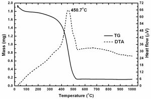 | Fig. 2- TG and DTA curves for the CHA. The value indicated represents an exothermic peak |
 | Fig. 3- XRD pattern for the CHA. Some peaks associated with crystalline phases can be observed |
 | Fig. 4- SEM images of the CHA. Magnifications: (a) 100x, (b) 500x, (c) 2000x, and (d) 5000x. Small pores can be observed on the rough surface at high magnification |
 | Table 1- Result of ICP-AES measurements of HA-C. Concentrations below the detection limit are marked as “*†|