


ISSN : 0976-7061
EISSN : 0976-707X



BAMASHMOUS S.A.1, AL-NUZAILY M.A.K.2, AL-MAKTARI L.A.S.3, TARESH S.A.G.4, ALI F.H.H.5*
1Haematology Department, Faculty of Medicine and Health Sciences, Sana’a University, Yemen.
2Haematology Department, Faculty of Medicine and Health Sciences, Sana’a University, Yemen.
3Haematology Department, Faculty of Medicine and Health Sciences, Sana’a University, Yemen.
4Haematology Department, Faculty of Medicine and Health Sciences, Sana’a University, Yemen.
5Haematology Department, Kuwait University Hospital, Sana’a University, Yemen.
* Corresponding Author : sshrmany@yahoo.co.uk
Received : 03-08-2013 Accepted : 31-08-2013 Published : 07-10-2013
Volume : 4 Issue : 1 Pages : 57 - 60
J Clin Res Lett 4.1 (2013):57-60
DOI : http://dx.doi.org/10.9735/0976-7061.4.1.57-60
Conflict of Interest : None declared
The aim of this study was to investigate the prevalence and etiology of anemia in hypothyroid women. In this study 120 recently diagnosed, non-treated hypothyroid women (overt=75, subclinical=45) and 60 healthy controls were included and subjected to determination of serum fT3, fT4, TSH, folic acid, vitamin B12, iron, iron-binding capacity and ferritin by Elecsys cobas analyzers, whereas complete blood count values by Sysmex XT-2000i automated hematology analyzer. Peripheral smears of the anemic patients were examined. The data was statistically analysed by SPSS-10 and p values less than 0.05 were considered significant. Our results showed that anemia prevalence was 48% in the overt hypothyroid group and 46.7% in the subclinical hypothyroid group and it was statistically meaningful compared to the control groups. Thus, the frequency of anemia in subclinical hypothyroidism is as high as that in overt hypothyroidism. There was no difference between the hypothyroid patients in terms of anemia. Folic acid, vitamin B12, and Fe were similar between these patients. Free T4 was significantly correlated with the erythrocyte indices )haemoglobin, hematocrit and MCV( (all P<0.05). TSH appeared not to be associated with any of the erythrocyte indices (all P>0.05). In conclusion, we confirmed that free T4 was associated with erythrocyte indices (Hb, PCV and MCV), confirming the role of thyroid hormones in the regulation of erythropoiesis. Also, anemia of chronic disease is the most common type of anemia in hypothyroid patients. Therefore, suspicion of hypothyroidism should be considered in anemias with uncertain etiology.
Anemia, Overt and Subclinical Hypothyroidism.
Although frequency of hypothyroidism differs from one society to another, 2-5% of prevalence has been reported through out the world. However, the prevalence of subclinical hypothyroidism is approximately 4-8.5%; it can reach to 20% in women aged 60 years or older [1] .
There is a metabolic deceleration in hypothyroidism. All organ systems are affected; and these symptoms and findings show different characteristics depending on the occurrence age of the hypothyroidism and deficiency or inefficacy of thyroid hormones. Hematopoietic system is the primary one among these affected systems and anemia is the most important one. Mediocre anemia is commonly seen in hypothyroidism. Anemia is defned in 20-60% of the patients with hypothyroidism [2,3] .
Anemia in hypothyroidism can be normochromic normocytic, hypochromic microcytic, and macrocytic. Anemia severity is associated with the hypothyroidism degree. Hypocellular structure of the bone marrow gives rise to thought that thyroid hormones play a role in hematopoiesis. The most frequently encountered anemia type is normochromic normocytic anemia. The most frequent reason of this is the bone marrow repression due to thyroid hormone defciency as well as lack of erythropoietin production arising from the reduction in need of O2. Erythrocyte life cycle in hypothyroidism is normal, and there is hypoproliferative erythropoiesis. Thyroid hormones also increase 2-3 DPG (diphosphoglycerate) levels assisting in the transmission of oxygen into the tissues [4-6] . Autoimmune thyroid disorders can be seen with other autoimmune disorders. Pernicious anemia can accompany hypothyroidism as a constituent of polyglandular autoimmune syndrome. Failure of vitamin B12 absorption occurs in pernicious anemia due to intrinsic factor (IF) deficiency and gastric achlorhydria.
This is the reason of macrocytic anemia occurrence in hypothyroidism. Macrocytosis is found in 55% of the hypothyroid patients [2] . Iron deficiency anemia is related with menorrhagia occurring as a result of various hormonal imbalances and also malabsorption which is seen in hypothyroidism [7,8] . Folic acid is another vitamin with impaired intestinal absorption, and causing macrocytic anemia in hypothyroidism [9] . In the present study we investigated the prevalence and etiology of anemia in hypothyroid women.
This study was conducted in Sana’a, Yemen, from April to July, 2013. It included 180 subjects aged 25 to 49 years. The patient’s group consisted of 120 recently diagnosed, non-treated hypothyroid women (overt=75, subclinical=45) (mean age ±SD, 36.7±5.84; median, 36.0; ranged from 28 to 49; 95% CI, 34.8-38.6 years old). These patients were selected randomly each day from subjects referred to the out-patient’s clinics of medical and general surgery departments (n=48) of Kuwait University Hospital (KUH) and from subjects referred to the specialized medical laboratories, Al-Aulaqi (n=42), Med-Lab. (ML) (n=21) and Al-Dubhani (n=9) for ELISA thyroid hormones measurements. The diagnosis of overt hypothyroidism was based on elevated serum TSH and decreased fT4 and/or fT3 levels, whereas the diagnosis of subclinical hypothyroidism was based on elevated serum TSH with normal fT4 and fT3 levels. The control group included 60 subjects (females) (mean age ±SD, 29.4±5.41; median, 28.5; ranged from 22-46; 95% CI, 26.8-32.0 years old) as normal healthy volunteers from the workers and students of KUH. All participants gave their informed consent to participate in this study.
Non-fasting blood samples five millilitres (5 ml) of venous blood were withdrawn under complete aseptic condition from each of patient and control group. From this 5 ml, 3 ml were put in plain tube and 2 ml in ethylenediaminetetraacetic acid (EDTA) tube. Sample of plain tube was left to clot for 30 minutes and serum was separated by centrifugation at 3500 x g for 5 minutes. Determination of fT3, fT4 and TSH concentrations were carried out immediately. The remaining serum samples were stored at -20°C for later analysis and estimation of folic acid, vitamin B12, iron, iron binding capacity and ferritin concentrations. EDTA samples were mixed well and estimation of complete blood count values were carried out immediately, then EDTA samples were stored at -20°C.
Measurements of serum concentrations of fT3, fT4, TSH, folate, vitamin B12 and ferritin were done using the electrochemiluminescence immunoassay (ECLIA) intended for use on the Elecsys reagent kits supplied by Roche Diagnostics GmbH (Mannheim, Germany) and run on cobas e 601immunoassay analyzer from Roche Diagnostics Ltd, Switzerland. The precision coefficient of variation (CV) of the method for fT3, fT4,TSH, folate, vitamin B12 and ferritin were 2.5%, 2.7%, 3.3%, 3.4%, 3.3% and 4.4%, respectively, and the reference ranges were 2.0-4.4 pg/mL, 0.93-1.7ng/dL, 0.27-4.20 mIU/mL, 10.4-42.4 nmol/L, 191-663 pg/mL and 13-150 µg/L, respectively.
Measurements of serum concentrations of iron and unsaturated iron-binding capacity (UIBC) were carried out by binding assay intended for use on the Elecsys reagent kits and run on Roche/Hitachi cobas c 501immunoassay analyzer. The precision CV of the method for iron and UIBC were 1.8% and 4.7%, respectively, and the reference ranges were 37-145 µg/dL and 112-346 µg/dL, respectively.
Estimation of complete blood count values were carried out by automated Sysmex XT-2000i automated hematology analyzer from Sysmex Corporation, (Kobe, Japan). Peripheral smears are done to confirm the type of anemia due to erythrocyte morphology and to exclude some other pathologies such as leukemia. Peripheral smears of anemic patients were examined.
Anemia is defined as hemoglobin levels lower than 11.2 g/dL in women. Iron deficiency anemia is defined as serum Fe levels lower than 37µg/dL, iron binding capacity greater than 346µg/dL, ferritin levels lower than 13µg/L and with microcytosis and hypochromia in peripheral blood smear. Folic acid deficiency anemia is defined as folic acid levels lower than 10.4nmol/L together with macrocystosis in peripheral blood smear. Vitamin B12 deficiency anemia is defined as B12 levels lower than 191pg/mL with increased MCV levels and with macrocystosis in peripheral blood smear. Anemia of chronic disease is defined as low Iron, low iron binding capacity and ferritin levels normal or elevated, folic acid and vitamin B12 levels normal.
All statistical analyses were performed using the Statistical Package for the Social Sciences (SPSS software ver. 10.0 for Windows, Inc., Chicago, Illions, USA) to indicate the degree of significant between the mean values of the patient groups and the mean values of the corresponding controls. Descriptive data were given as mean ± standard deviation (SD). All tests were two-tailed and p values less than 0.05 were considered statistically significant. Pearson correlation test was used in correlations between parametric variables.
In our study; Anemia was present in 57 of 120 hypothyroid patients included into the study. Anemia frequency was 47.5% in the hypothyroid patient groups and it was statistically meaningful compared to the control group (p=0.001). Anemia was determined in 48% of those with overt hypothyroidism and 46.7% of those with subclinical hypothyroidism [Table-1] and it was statistically meaningful compared to the control group (p=0.001). Biochemical data of the patients with anemia in hypothyroid and control groups are shown at [Table-2] .
There was no statistical difference between ferritin levels of the anemic patients and control groups [Table-2] . The etiologies of anemia in patients and controls are shown in [Table-3] .
In overt and subclinical hypothyroid patient groups, anemia of chronic disease frequency was found to be the most common type. It was present in 36 of 120 (30%) of patient groups. The frequencies of iron deficiency anemias, folic acid deficiency and vitamin B12 deficiency were 7.3%, 5.35% and 5.35% respectively.
In the subgroup analysis of the hypothyroid patients, the most frequently seen anemia type was the anemia of chronic disease in patients with overt and subclinical hypothyroidism. Anemia of chronic disease was determined in 24 of 75 (32%) patients with overt hypothyroidism, and in 12 of 45 (26.7%) patients with subclinical hypothyroidism.
There was microcytic anemia in 6 of 75 (8%) overt hypothyroid patients and in 3 of 45 (6.6%) subclinical hypothyroid patients. There was macrocytic anemia in 6 of 75 (8%) overt hypothyroid patients and in 6 of 45 (13.3%) subclinical hypothyroid patients. There was normocytic anemia in 32% of overt hypothyroid patients and in 26.7% of subclinical hypothyroid patients.
Free T4 was significantly associated with the all erythrocyte indices (haemoglobin, hematocrit and MCV (p=0.001, p=0.001 and p=0.004, respectively). TSH appeared not to be associated with any of the erythrocyte indices (all P>0.05).
According to the data of WHO, anemia prevalence is 24.8% throughout the world and it is seen more frequently in underdeveloped countries [10] . In our study, we observed that anemia frequency in overt and subclinical hypothyroid groups was determined to be 48% and 46.7%, respectively. Anemia frequency in patients with overt and subclinical hypothyroidism was found to be statistically significant when compared to control group (p=0.001). This result gave rise to thought that hypothyroidism presence may be a risk factor in anemia development.
Directly or indirectly, stimulation of erythroid colony development by thyroid hormones, inhibition of the latter in its absence, reduction in oxygen distribution to tissues and diminution of erythropoietin level in the absence of thyroid hormones causes normocytic anemia that this anemia forms the most frequent type of anemia in hypothyroid patients [11] . The determination made by Christ-Crain, et al [3] indicated that erythropoietin values were increased as result of levothyroxine treatment in women with subclinical hypothyroidism. Also in our study, similar with the literature the most frequent anemia type was anemia of chronic disease in overt and subclinical hypothyroid groups. However, prevalence of vitamin B12 deficiency increases along with the age and Framingham study reveals that the prevalence in old population is 12% [12] . Wang, et al [13] found that vitamin B12 deficiency was 19.71% and megaloblastic anemia prevalence was 9.82% in old hospitalized patients in the neurology clinic. However, the prevalence is observed as 1.6% to 10% in Europe [14] . In our study, we found that vitamin B12 deficiency was 5.35% in hypothyroid groups. Another reason of anemia is macrocytic anemia occurring as a result of vitamin B12 deficiency. It mostly occurs as a result of malabsorption due to pernicious anemia accompanying hypothyroidism. Antibodies against gastric parietal cells were determined in 1/3 of the patients with primary hypothyroidism [15] . It was also determined that the presence of clinical pernicious anemia was seen in 10% of the patients [16,17] . In the study carried out by Carnel, et al [18] , thyroid disorder and hypothyroidism were determined respectively in 24.1% and 11.7% of the patients with pernicious anemia. Insufficient intake, absorption change arising from deceleration in intestinal motility, intestinal wall oedema, and bacterial infiltration are blamed among other reasons causing vitamin B12 deficiency in hypothyroidism [16] . Jabbar, et al [16] evaluated the prevalence of vitamin B12 deficiency and found low vitamin B12 levels in 46 of 116 (39.6%) patients consulted to endocrinology department.
Folic acid deficiency, one of the reasons of anemia, occurs as a result of intestinal malabsorption. Again hypothyroidism ruins folate mechanism by decreasing hepatic level of dihydrofolate reductase such as methylenetetrahydrofolate reductase [19] . Folic acid deficiency almost always occurs as secondary to an underlying disease. We determined folic acid deficiency in hypothyroid patients as 5.3% in our study.
One of the most frequently seen diseases in all over the world is iron deficiency anemia. In a study carried out in England, it was reported that its frequency was between 3.5-5.3% [20] . In our study, iron deficiency anemia frequency was 7.3% in hypothyroid patients. Malabsroption and iron deficiency anemia depending on menorrhagia occurring as a result of various hormonal instability are observed in hypothyroidism. In a study carried out by Cinemre, et al [8] they showed that the efficacy and absorption of oral iron treatment in women with subclinical hypothyroidism improved after levothyroxine replacement. This demonstrates that hypothyroidism should be assessed in patients with anemia.
Hypothyroidism is an endocrine disorder that is frequently seen in the society. Anemia is frequently seen in these patients and it occurs as a result of various causes. Determination of etiological reasons of anemia and arrangement of the treatment is important. As a result, we found an elevated anemia frequency in hypothyroid patients consistent with the literature. We determined that the most frequent cause was linked to anemia of chronic disease. As a result increase of anemia frequency in hypothyroidism is detected in our study. Further studies with larger number of patients are needed to clarify this increase.
In conclusion, we confirmed that free T4 was associated with erythrocyte indices (Hb, PCV and MCV), confirming the role of thyroid hormones in the regulation of erythropoiesis. Also, anemia of chronic disease is the most common type of anemia in hypothyroid patients. Therefore, suspicion of hypothyroidism should be considered in anemias with uncertain etiology.
The authors are grateful to all doctors of Lab. and the health workers in the KUH and specialized medical laboratories, Al-Aulaqi, Med-Lab. and Al-Dubhani for their help and cooperation during this work.
[1] Wilson G.R. and Curry R.W. (2005) Am. Fam. Physician, 72(8), 1517-1524.
» CrossRef » Google Scholar » PubMed » DOAJ » CAS » Scopus
[2] Antonijević N., Nesović M., Trbojević B., Milosević R. (1999) Med. Pregl., 52(3-5), 136-140.
» CrossRef » Google Scholar » PubMed » DOAJ » CAS » Scopus
[3] Christ-Crain M., Meier C., Huber P., Zulewski H., Staub J.J., Müller B. (2003) Hormones, 2, 237-242.
» CrossRef » Google Scholar » PubMed » DOAJ » CAS » Scopus
[4] Horton L., Coburn R.J., England J.M., Himsworth R.L. (1976) Q. J. Med., 45(177), 101-123.
» CrossRef » Google Scholar » PubMed » DOAJ » CAS » Scopus
[5] Das K.C., Mukherjee M., Sarkar T.K., Dash R.J., Rastogi G.K. (1975) J. Clin. Endocrinol. Metab., 40, 211-220.
» CrossRef » Google Scholar » PubMed » DOAJ » CAS » Scopus
[6] Fein H.G., Rivlin R.S. (1975) Med. Clin. North Am., 59, 1133-1145.
» CrossRef » Google Scholar » PubMed » DOAJ » CAS » Scopus
[7] Lawrence E., Shapiro A., Surks M.I. (2001) Principles and Practice of Endocrinology and Metabolism, 3rd ed. Lippincott Williams & Wilkins, Philadelphia, 445-451.
» CrossRef » Google Scholar » PubMed » DOAJ » CAS » Scopus
[8] Cinemre H., Bilir C., Gokosmanoglu F., Bahcebasi T. (2009) J. Clin. Endocrinol. Metab., 94(1), 151-156.
» CrossRef » Google Scholar » PubMed » DOAJ » CAS » Scopus
[9] Sims E.G. (1983) J. Natl. Med. Assoc., 75(4), 429-431.
» CrossRef » Google Scholar » PubMed » DOAJ » CAS » Scopus
[10] Benoist B., McLean E., Egli I., Cogswell M. (2008) WHO Global Database on Anemia, Geneva, 1-2.
» CrossRef » Google Scholar » PubMed » DOAJ » CAS » Scopus
[11] Kuhr T., Hala K., Dietrich H., Herold M., Wick G. (1994) J. Autoimmune, 7, 13-25.
» CrossRef » Google Scholar » PubMed » DOAJ » CAS » Scopus
[12] Lindenbaum J., Rosenberg I.H., Wilson P.W., Stabler S.P., Allen R.H. (1994) Am. J. Clin. Nutr., 60(1), 2-11.
» CrossRef » Google Scholar » PubMed » DOAJ » CAS » Scopus
[13] Wang Y.H., Yan F., Zhang W.B., Ye G., Zheng Y.Y., Zhang X.H., Shao F.Y. (2009) Neuroscience Bulletin, 25(4), 209-215.
» CrossRef » Google Scholar » PubMed » DOAJ » CAS » Scopus
[14] McLean E., de Benoist B., Allen L.H. (2008) Food Nutr. Bull., 29(2), S38-51.
» CrossRef » Google Scholar » PubMed » DOAJ » CAS » Scopus
[15] Mehmet E., Aybike K., Ganıdagli S., Mustafa K. (2012) Endocrine Journal, 59(3), 213-220.
» CrossRef » Google Scholar » PubMed » DOAJ » CAS » Scopus
[16] Jabbar A., Yawar A., Waseem S., Islam N., Ul Haque N., Zuberi L., Khan A., Akhter J. (2008) Journal of the Pakistan Medical Association, 58(5), 258-261.
» CrossRef » Google Scholar » PubMed » DOAJ » CAS » Scopus
[17] Kaferle J. and Strzoda C.E. (2009) Am. Fam. Physician, 79(3), 203-208.
» CrossRef » Google Scholar » PubMed » DOAJ » CAS » Scopus
[18] Carmel R. and Speneer C.A. (1982) Arch. Intern. Med., 142(8), 1465-1469.
» CrossRef » Google Scholar » PubMed » DOAJ » CAS » Scopus
[19] Das K.C., Mukherjee M., Sarkar T.K., Dash R.J., Rastogi G.K. J. Clin. Endocrinol. Metab., 40(2), 211-220.
» CrossRef » Google Scholar » PubMed » DOAJ » CAS » Scopus
[20] Mukohopadhyay D., Mohannaruban K. (2002) Age Ageing, 31(2), 87-91.
» CrossRef » Google Scholar » PubMed » DOAJ » CAS » Scopus
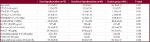 | Table 1- Clinical and laboratory results of patients groups and control group |
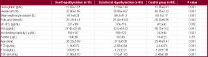 | Table 2- Laboratory results of the anemic patients groups and control group |
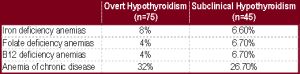 | Table 3- Etiology and percentages of anemia in overt and subclinical hypothyroidism |