


ISSN : 2321-4422
EISSN :



SAWANT S.P.1*
1Department of Anatomy, K. J. Somaiya Medical College, Somaiya Ayurvihar, Eastern Express Highway, Sion, Mumbai- 400 022, MS, India.
* Corresponding Author : drspsawant@gmail.com
Received : 04-01-2013 Accepted : 14-02-2013 Published : 01-04-2013
Volume : 1 Issue : 1 Pages : 1 - 3
World Res J Cardiol 1.1 (2013):1-3
Conflict of Interest : None declared
During routine dissection, of the right upper limb of a 70 years old donated embalmed male cadaver in the Department of Anatomy, K.J. Somaiya Medical College, Sion, Mumbai, India, we observed a trifurcation of the brachial artery into the radial, ulnar and common interosseous arteries. The brachial artery trifurcated above the cubital fossa in the lower part of the arm. The common interosseous artery was longer in length. The common interosseous artery divided at an unusual site in the cubital fossa into the anterior and posterior interosseous arteries. The radial artery travelled downward along the radial side of the forearm to the wrist. The course of the ulnar artery was normal. There were no associated altered anatomy of the nerves observed in the specimen. The variation was unilateral and the left upper limb was normal. The photographs of the trifurcation of brachial artery into radial, ulnar and common interosseous artery were taken for proper documentation. <b>Conclusion-</b> Topographical anatomy of the normal and abnormal variations of the axillary artery are clinically important for surgeons, orthopaedicians operating on the supracondylar fracture of humerus and radiologists performing angiographic studies on the upper limb. The trifurcation of brachial artery in the lower part of arm may result in excessive haemorrhage during supracondylar fracture of the humerus. A lack of knowledge of such type of variations with different patterns may complicate the surgery and may cause unnecessary bleeding. Therefore both the normal and abnormal anatomy of the region should be well known for accurate diagnostic interpretation and therapeutic intervention.
Brachial Artery, Trifurcation, Radial Artery, Ulnar Artery, Common Interosseous Artery, Surgeons, Orthopaedicians, Supracondylar Fracture, Radiologists, Angiographic Studies.
The brachial artery ends in the cubital fossa by dividing into the radial and ulnar arteries. The ulnar artery, the deeper and the larger of the two terminal branches of the brachial artery, begins a little below the bend of the elbow, and, passing obliquely downward, reaches the flexor carpi ulnaris muscle in its middle third, whereas the ulnar nerve is covered by the muscle throughout its entire course running under the tendon in the wrist region.It then runs along the ulnar border upto the wrist, crosses the transverse carpal ligament on the radial side of the pisiform bone, and immediately beyond this bone divides into two branches, which enter into the formation of the superficial and deep palmar arches. The common interosseous artery is a short branch of the ulnar, about 1 cm. in length, arises immediately below the tuberosity of the radius from the Ulnar artery. It passes back to the upper border of the interosseous membrane and divides into anterior and posterior interosseous arteries. Anterior interosseous artery descends on the anterior aspect of the interosseous membrane with the median nerve's anterior interosseous branch. Median artery, a slender branch from anterior interosseous artery, accompanies and supplies the median nerve [1] . The radial artery appears, from its direction, to be the continuation of the brachial, but it is smaller in caliber than the ulnar. It commences at the bifurcation of the brachial, just below the bend of the elbow, and passes along the radial side of the forearm to the wrist and take part in the completion of the superficial and deep palmar arches.
During routine dissection, of the right upper limb of a 70 years old donated embalmed male cadaver in the Department of Anatomy, K.J. Somaiya Medical College, Sion, Mumbai, India, we observed a trifurcation of the brachial artery into the radial, ulnar and common interosseous arteries. The brachial artery trifurcated above the cubital fossa in the lower part of the arm. The common interosseous artery was longer in length. The common interosseous artery divided at an unusual site in the cubital fossa into the anterior and posterior interosseous arteries. The radial artery travelled downward along the radial side of the forearm to the wrist. The course of the ulnar artery was normal. There were no associated altered anatomy of the nerves observed in the specimen. The variation was unilateral and the left upper limb was normal. The photographs of the trifurcation of brachial artery into radial, ulnar and common interosseous artery were taken for proper documentation.
The brachial artery commonly terminates into radial and ulnar arteries proximal or distal to intercondylar line. But the trifurcation of the brachial artery into radial, ulnar and common interosseous arteries are not common [1] . In the present case the brachial artery trifurcates into the radial, ulnar and common interosseous arteries above the supracondylar line in the lower part of the arm. The supernumerary branches of brachial artery may be the radial recurrent artery or the median artery [2] . Various authors have made studies on termination of brachial artery [1-10] . It may bifurcate proximally and reunite to form single trunk. Sometimes ulnar artery arise proximally. Rarely there may be a communicating vessel connecting axillary artery and brachial artery [1] . The radial recurrent arising from the lower part of brachial artery separately but not as a one of the terminal branch is reported in literature [2] . The trifurcation of brachial artery into ulnar, radial, and radial recurrent arteries in a right superior extremity of fifty years old male cadaver during dissection is documented in literature. The third branch was radial recurrent artery and the common interosseous artery was given off from the ulnar artery, which divided into anterior and posterior interosseous arteries. The radial artery was normal in that study [10] . In the present case, the brachial artery had trifurcated into ulnar, radial, and common interosseous arteries in the right upper limb. There were no communicating branches seen between radial and ulnar arteries. In high termination of brachial artery if one of the two arteries lies superficial to the superficial flexor group of muscles. The other artery is taking the usual course is crossed superficially by the median nerve [11] . In the present case the ulnar artery was present deep to the superficial flexor muscles of forearm and no aberrant artery was observed. The ulnar artery may take origin from the brachial artery proximally and then the brachial artery terminates into the radial artery and the common interrosseous artery in the cubital fossa [16] . The radial artery also may take origin proximally from the brachial artery running superficial to forearm flexors or deep fascia or rarely subcutaneous. The common interosseous artery may take origin proximally [17] . Sometimes the radial artery may be absent [18] and even the brachial artery may be absent [19] .
The seventh cervical intersegmental artery forms the axis artery of the upper limb and persists in the adult to form the axillary, brachial, and interosseous arteries. Transiently, the median artery arises as a branch of the interosseous artery, begins to regress and remains as a residual artery accompanying the median nerve [13] . The radial and the ulnar arteries are later additions to the axis artery. The ulnar artery and the median artery are branches of the axis artery [12] . The superficial brachial artery is a consistent embryonic vessel, coexisting or not with the brachial artery [14] . It has two terminal branches, lateral and medial. The lateral continues as a part of the definitive radial artery [15] and the medial i. e. superficial antebrachial artery, which divides into the median and ulnar artery branches, which are the trunks of origin of the median and ulnar arteries. The arterial pattern of the upper limb develops from an initial capillary plexus by a proximal and distal differentiation, due to maintenance, enlargement and differentiation of certain capillary vessels, and the regression of others. The number of upper limb arterial variations arise through the persistence, enlargement and differentiation of parts of the initial network which would normally remain as capillaries or even regress [12,20-22] .
The knowledge of presence of the unusual high level trifurcation of brachial artery is clinically important for clinicians, surgeons, orthopaedicians and radiologists performing angiographic studies. Undoubtedly, such variations are important for diagnostic evaluation and surgical management of vascular diseases and injuries.
The trifurcation of brachial artery in the lower part of arm may result in excessive haemorrhage during supracondylar fracture of the humerus. A lack of knowledge of such type of variations with different patterns may complicate the surgery and may cause unnecessary bleeding. Therefore both the normal and abnormal anatomy of the region should be well known for accurate diagnostic interpretation and therapeutic intervention.
The author declare that he has no competing interests.
Author is thankful to the Dean, Dr. Geeta Niyogi for her support and encouragement. I would like to thank Mr. M. Murugan, Mr. Sanjay, Mr. Pandu, Mr. Kishor, Mr. Nitin, Mr. Sankush and Mr. Raju for their help. Authors also acknowledge the immense help received from the scholars whose articles are cited and included in references of this manuscript. The author is also grateful to authors / editors / publishers of all those articles, journals and books from where the literature for this article has been reviewed and discussed.
[1] Williams P.L., Bannister L.H., Berry M.M. Colins P., Dyson M., Dussek J.E. and Ferguson M.W.J. (2005) Gray’s Anatomy, 39th ed., Churchill Livingstone, New York, 939.
» CrossRef » Google Scholar » PubMed » DOAJ » CAS » Scopus
[2] Huber G.C., (1930) The Vascular System. 9th ed., J.B. Lippincott Co., Philadelphia, 767-791.
» CrossRef » Google Scholar » PubMed » DOAJ » CAS » Scopus
[3] Massie G. (1944) Surgical Anatomy, 4th ed., J & A Churchill Ltd, London, 136-139.
» CrossRef » Google Scholar » PubMed » DOAJ » CAS » Scopus
[4] Thorek P. (1951) Anatomy in Surgery, 2nd ed., J.B. Lippincott Co, Philadelphia, 702-8.
» CrossRef » Google Scholar » PubMed » DOAJ » CAS » Scopus
[5] Anson B.J. and Maddock W.G. (1952) Callander’s Surgical Anatomy, 3rd ed. W.B. Saunders Co., Philadelphia, 762-764.
» CrossRef » Google Scholar » PubMed » DOAJ » CAS » Scopus
[6] Anson B.J. (1966) Morris’ Human Anatomy, McGraw Hill Book Co., The Blackiston Division, New York, 708-724.
» CrossRef » Google Scholar » PubMed » DOAJ » CAS » Scopus
[7] Boyd J.D., Clark W.E., Hamilton W.J., Yoffey J.M., Zuckerman S and Appleton A.B. (1956) Textbook of Human Anatomy, MacMillan and Co. Ltd. St. Martin’s Press, New York, 341-346.
» CrossRef » Google Scholar » PubMed » DOAJ » CAS » Scopus
[8] Lockhardt R.D., Hamilton G.F. and Fyfe F.W. (1959) Anatomy of the Human Body, Faber and Faber Limited, London, 612-619.
» CrossRef » Google Scholar » PubMed » DOAJ » CAS » Scopus
[9] Romanes G.J. (1644) Cunningham’s Textbook of Anatomy, 10th ed., Oxford University Press, New York, 885-893.
» CrossRef » Google Scholar » PubMed » DOAJ » CAS » Scopus
[10] Patnaik V.V.G., Kalsey G., Singla Rajan (2001) Journal of Anatomical Society of India, 50(2), 163-165.
» CrossRef » Google Scholar » PubMed » DOAJ » CAS » Scopus
[11] Hollinshed Henry (1962) Anatomy for Surgeons-Back and Limbs, 2, 4th ed., 214.
» CrossRef » Google Scholar » PubMed » DOAJ » CAS » Scopus
[12] Rodriguez-Baeza A., Nebot J., Ferreira B., Reina F., Perez J., Sanudo J.R. and Rolg M. (1995) Journal of Anatomy, 187, 473-479.
» CrossRef » Google Scholar » PubMed » DOAJ » CAS » Scopus
[13] Singer E. (1933) Anatomical Record, 55, 403-409.
» CrossRef » Google Scholar » PubMed » DOAJ » CAS » Scopus
[14] Tountas CH.P. and Bergman R.A. (1993) Anatomic Variations of the Upper Extremity, Churchill Livingstone, New York, 196-210.
» CrossRef » Google Scholar » PubMed » DOAJ » CAS » Scopus
[15] Vancov V. (1961) Anatomischer Anzeiger, 109, 400-405.
» CrossRef » Google Scholar » PubMed » DOAJ » CAS » Scopus
[16] Aharinejad S., Nourani F. and Hollensteiner H. (1997) Clinical Anatomy, 10, 253-258.
» CrossRef » Google Scholar » PubMed » DOAJ » CAS » Scopus
[17] Celik H.H., Germus G., Aldur M.M. and Ozcelik M. (2001) Morphologie, 85, 25-27.
» CrossRef » Google Scholar » PubMed » DOAJ » CAS » Scopus
[18] Suganthy J., Koshy S., Indrasingh I. and Vettivel S. (2002) Journal of the Anatomical Society of India, 51(1), 61-64.
» CrossRef » Google Scholar » PubMed » DOAJ » CAS » Scopus
[19] Clerve A., Kahn M., Pangilinan A.J. and Dardik H. (2001) Journal of Vascular Surgery, 33, 191-194.
» CrossRef » Google Scholar » PubMed » DOAJ » CAS » Scopus
[20] Chummy S.S. (2001) Last’s Anatomy Regional and Applied, 10th ed., New York, Churchill Livingston, 48-55.
» CrossRef » Google Scholar » PubMed » DOAJ » CAS » Scopus
[21] Moore K.L. and Persand T.V.N. (2003) Clinically Oriented Embryology, 7th ed., Elsevier, India, 329-80.
» CrossRef » Google Scholar » PubMed » DOAJ » CAS » Scopus
[22] Hamilton W.J. and Mossman H.W. (1978) Human Embryology, 4th ed., Baltimore, Williams &Wilkins, 268-72.
» CrossRef » Google Scholar » PubMed » DOAJ » CAS » Scopus
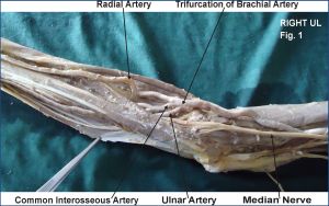 | Fig. 1- The photographic presentation of the variant trifurcation of brachial artery into radial, ulnar and common interosseous artery. |
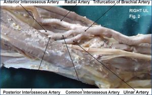 | Fig. 2- The photographic presentation of unusual termination of the common interosseous artery into the anterior and posterior interosseous arteries in the cubital fossa. |