


ISSN : 2321-4414
EISSN :



HEDAYAT A.1*, ELMOSELHI H.2, SHOKER A.3
1College of Dentistry, University of Saskatchewan, SK, Canada S7N 5E4.
2Saskatchewan Transplant Program, St. Paul’s Hospital, Saskatoon, SK, Canada S7M 0Z9.
3Department of Medicine, Division of Nephrology, University of Saskatchewan, 103 Hospital Dr., Saskatoon, SK, Canada S7N 0W8.
* Corresponding Author : assem.hedayat@usask.ca
Received : 06-01-2013 Accepted : 07-03-2013 Published : 01-04-2013
Volume : 1 Issue : 1 Pages : 1 - 9
World Res J Nephrol 1.1 (2013):1-9
Conflict of Interest : None declared
Historically, most models explaining and predicting the transport of uremic toxins through hemodialysis membranes focused on the molecular kinetics mechanism as dictated by both the concentration and barometric gradients. The design and permselectivity of hemodialysis membranes revolved around the concentration and the molecular weight of the uremic toxins. With the emerging nanotechnology, and our capability to nanofabricate thinner hemodialysis membranes with nanopores of unique geometrical configurations and sizes, non-equilibrium (irreversible) thermodynamics will dominate the clearance of uremic toxins through the nanofabricated membranes, and new design parameters such as molecular size, shape, electric charges, and molecular conformity will be considered as sieving parameters. We catalogued these properties for some uremic toxins. Designing and nanofabricating efficient hemodialysis membranes is subject to our success in bringing the three main driving forces of molecular sieving into synergy. These driving forces are: diffusion (concentration gradient), convection (pressure gradient), and migration (electric potential gradient). This will lead to closing the wide gap between the filtration of a naturally functioning kidney and artificial hemodialysis membranes. Other requirements of successful nanofabricated membranes are: biocompatibility and non-thrombogenicicity, and optimal balance between hydrophobicity and hydrophilicity. A nanofabricated hemodialysis membrane with all these characteristics will improve quality of life, morbidity, mortality, hemodialysis standards for ESRD patients, and will be cost effective for global economy.
Hemodialysis, Flux, Toxins, Clearance, Nanotechnology
Nanofabricated hemodialysis membranes.
Recent technological advancement in engineering, science, and medicine is clearly reflected in the technical terms we use. As recent as the 1990’s, we used the terms: microtechnology, and micropores. In the last decade, these terms were transformed into: nanotechnology, and nanopores, and a number of facilities, laboratories, and businesses around the world replaced their activity description of microfabrication by nanofabrication.
Nanotechnology is the technology of producing nanoscale (10-9 m) systems with dimensions in the range of 1-100 nm [1] . In medicine, a new subdivision: nanomedicine has emerged. This health science deals with structures that are a thousand times smaller than a cell, and is predicted to improve the effectiveness of medicine since it can tackle disease, one cell at a time [2] . It has great potential in early detection of disease, and consequently, in increasing the probability of patients’ survival [2] . Within nanomedicine, further new subdivisions consequently emerged: nanoophthalmology [3] , and nanonephrology [1] . This article will focus on one aspect of nanonephrology, which is the nanofabrication of hemodialysis membranes.
Uremic toxins have different molecular weights, shapes, sizes, conformations, and can be either neutral or with net positive or negative charges on them. Urea and creatinine are small, hydrophilic molecules [4] , with molecular weights of 60 and 113 Da, respectively. Albumin has a large molecular weight at 69,000 Da. Both β2-Microglobulin and Vitamin B12 belong to middle molecules with molecular weights of 11,800 and 1,355 Da, respectively [5] .
Albumin has a molecular radius of gyration of 27.4 +/- 0.35 Aº. It can have the form of a non-symmetrical oblate ellipsoid with a diameter of 85 Aº, and it can be an extended ellipsoid, 110 Aº in length [6] . Molecular shapes of molecules can be altered in the presence of external forces, such as an electric field [7] .
Human renal clearance processes are glomerular filtration, active tubular secretion and passive tubular reabsorption [8] . Renal clearance is mainly governed by physicochemical determinants such as: the molecular ionization state, lipophilicity, and polar descriptors. The permeability of renal membranes to molecules is decided by the ionization state of the molecules. However, high permeability of a membrane does not mean clearance of the molecules. Significant reabsorption may lead to minimize their clearance [9] .
In a glomerulus, the pore shape is a slit, 4.5-5 nm in size on average. In glomerular filtration, molecules with a hydrodynamic diameter of less than 6nm are usually filtered. For molecules with a hydrodynamic diameter between 6 and 8 nm, the filtration depends on the molecular size and net charge on it. Studies showed that glomerular filtration for similar sized molecules is greatest for cationic molecules, followed by neutral ones, followed by anionic ones.
Historically, regenerated cellulose was the first permeable membrane used in hemodialysis in the 1940’s. The next development was the use of hollow fiber hemodializers, which became popular in the 1970’s. By the mid 1970’s, the membrane’s thickness was reduced from 16 μm to 8 μm, and by the mid 1980’s, new synthetic materials, microgeometries, and morphologies were introduced as developments of hemodialysis membranes. This was followed by the use of high-flux membranes that have their drawbacks [10] .
The available hemodialysis technology does not provide ESRD patients with an acceptable quality of life. The morbidity and mortality rates of these patients remain high [11] .
Current hemodialysis focuses the sieving function on molecular weight exclusion, and the gap between the functional performance of a natural kidney and hemodialysis is vastly wide [12] . Commercially available hemodialysis membranes differ from one another with respect to the degree of bioincompatibility, chemical composition, structure, and transport characteristics. Cellulose membranes, for example are very hydrophilic. They allow small molecules (< 300 Da) to pass through but not the middle sized ones (300 to 12,000 Da). Other thermoplastic, synthetic membranes such as polysulphones and polyamides are hydrophobic, and are less restrictive to middle and large molecules. The higher the hydrophobicity of the membrane is, the higher will be its adsorbtive capacity to proteins, which is undesireable [13] .
Some examples of currently used hemodialysis membranes are HemophanR with a hollow fiber pore size distribution of 12 to 15 nm, and a 20% volume porosity. At 310ºK, the diffusivities of creatinine and vitamin B12 through the membrane are: 1.29 x 10⁻⁹ and 0.379 x 10⁻⁹ m2 sec-1, respectively. Cuprophan membranes have a varying pore radius of 2 to 3 nm, and a surface porosity of 30%. Finally, cellulose membranes vary in pore radii from 3 to 25 nm [14] . The variations in surface areas and thicknesses of low and high flux hemodialysis membranes are summarized in [Table-1] [15] . Low-flux and high-flux membranes produced by the same manufacturer and with the same surface areas (1.4 m2) have a pore radius of 3.1 and 5.7 nm, respectively [17] .
Molecular transport across a hemodialysis membrane can occur by one of two processes or driving forces: Diffusion as a result of a difference in solute concentration driving force where solutes move from high to low concentration. The transport of smaller solutes is rapid, and the rate decreases with the increase in molecular size. The diffusion of the solute depends upon the thickness of the membrane and its porosity. The second transport process is convection, where hydraulic or osmotic pressure differences across the membranes are the driving force for the trans-membrane fluid movement. The flux rate is directly proportional to the rate of fluid movement across the membrane, and the sieving coefficient is the proportionality constant between the rates of movement of solute and fluid [17] .
If a flexible molecule enters a pore that has a smaller diameter than its radius of gyration, the molecule will try to stretch itself by exerting energy to overcome an entropic energy barrier. It can get trapped at the pore’s interface. This is known as entropic trapping. This can occur even if the pore’s size is much larger than the backbone radius of the flexible molecule [18] .
Quantitatively, Kt/V is a dimensionless number that quantifies the adequacy of hemodialysis treatment, where K is the dialyzer’s clearance of urea (ml/min), t is the dialysis treatment time (min), and V is the urea distribution volume (ml). The Kt/V number proved to have an effect on morbidity and mortality of ESRD patients [19] . However, Kt/V quantification is limited in its usefulness by two factors: It only addresses the removal of small solutes such as urea, and the protocols of dialysis treatment vary from one country to the other [12] .
For a molecule to pass through a filter membrane of a specific porosity, the pore’s size should be larger than the largest dimension of the molecule designated to pass through. If the molecules do not pass through the pore freely, overlapping molecular conformations will occur leading to a lower degree of entropy for the molecules inside the membrane. Consequently, the coefficient of particle distribution between the pore and the solution adjacent to it will decrease, yielding a drop in the membrane’s permeability [7] .
Also, applied fields such as the electrical fields on the surface of the membrane can add to the complexity of filtration. For example, some molecules can change shape in the presence of electrical fields [7] .
The hindered transport theory explains the interaction between molecules and membrane pores. Firstly, molecular sieving can be affected by steric hindrance, where, statistically, it is harder for a larger molecule to fit in a pore than it is for a smaller molecule (steric partitioning). Also participating in hindrance is the Debye repulsion layer that exists when both pore wall and molecules are similarly charged (electrostatic partitioning). Secondly, it can be affected by hydraulic hindrance where the molecular motion in a pore is slowed down by the presence of adjacent static walls. This gives rise to drag forces on the molecules and slows them down. Both steric and hydraulic hindrance affects both diffusion and convection of the molecules through the pores of the membrane [18] .
From the developed mathematical models in the literature, we can deduce that the solute transport rate across a hemodialysis membrane is proportional to the membrane’s porosity, and the sieving coefficient of the membrane is inversely proportional to the thickness of the membrane [15] .
The objective of hemodialysis is to remove uremic toxins including middle molecules from the blood. Among the known toxins, 68 compounds have a molecular weight less than 500 Da, and 12 compounds exceed 12,000 Da. In general, middle molecules are a category of uremic toxins with molecular weights in the range of 300 to 12,000 Da [20] .
Proteomic analysis of hemodialysis fluids for humans yielded interesting statistics: More than half of the proteins had molecular weights less than 30,000 Da, and 85% were less than 60,000 Da. Also, most of the human proteins have molecular weights between 15,000 Da and 30,000 Da [5] . [Table-2] shows classical clearance of well-known molecules by the current hemodialysis membranes.
The clearance of a membrane to a specific molecule can be estimated through mathematical models. For example, to calculate the membrane’s clearance, Kd, of β2-Microglobulin the [Eq-1] was used:
Where, C(0) and C(T) are the predialysis and postdialysis β2-Microglobulin plasma concentrations, respectively. Qf is the average rate of ultrafiltration, T is the duration of session, and V is the extracellular fluid.
Values for Kd were compared to the calculated ones using the concentration differences between the molecule’s arterial and venous concentration difference across the dialyser. The model yielded membrane clearance values in the range of 54.4 to 74.4 ml/min. While the values agreed for low-flux membranes, they differed for high-flux ones. This means that other factors affect the clearance of β2-Microglobulin in high-flux dialysers [17] .
Although the literature reports the concentrations of uremic toxins as well as their molecular weight, little is known about their sizes, shapes, orientations, conformations, and the neutrality or net electric charges associated with each of them. These parameters are indispensable in the design and nanofabrication of hemodialysis membranes.
Polymeric membranes are not considered the ideal material to be used in hemodialysis. They are a poor representation of the glomerular filtration barrier because of the following reasons:
Hemodialysis membranes used by end stage renal disease patients [ESRD] have numerous weaknesses. They are made of polymeric materials such as cellulose acetate, polyethylene polyvinyl alcohol, polymethyl methacrylate, and polyether sulphone [13] . During hemodialysis, contact between the blood and the artificial membrane surface leads to a variety of interactions. These interactions include coagulation, platelet, leukocytes and compliment activation, cytokine production and production of free oxygen radicals among other events known as
bioincompatibility of hemodialysis membranes. These reactions cause injury to patients. Most of the incompatibility occurs because of the different characteristics of the membranes. It should be known also that bioincompatibility occurs due to the flow of blood through the extracorporeal circuit and the type of patient being treated. Bioincompatibility has long been considered as a main problem in dialysis treatment [27-42] .
Bioincompatibility causes inflammation in dialysis patients and therefore affects their morbidity and mortality. For example, the cardiac effects of chronic inflammation in dialysis patients are well recognized. The prevalence of cardiac disease is high in uremic patients just beginning dialysis and even more so in cases of late referral. The excessive risk of cardiac diseases in chronic uremic patients is in part due to dialysis related bioincompatibility [43] .
Within the hemodialysis membrane, the pores vary in size and density [13] , and the broad pore size distribution, inconsistent pore shape, and low transport rate lead to low dialysis efficiency [44] . For a large dialyzer, the surface area of the membrane exceeds 2 m2, and the membrane’s thickness is about 8 μm. Such a membrane thickness forms a diffusion barrier to some solutes. Also, the flow rate of middle sized toxins such as Beta2-microglobulin (11,800 Da) becomes very low (<10 ml / min or zero) when passing through low -permeability membranes. If high-flux membranes are used, middle-sized molecules pass through the membrane at a higher rate (>20 ml/min), but also beneficial molecules, such as albumin (60,000 Da), undesirably pass through [13] . In addition, polymeric membranes cause greater loss of protein due to adsorption, and their higher hydraulic permeability may increase the risk of back filtration and contamination [45] .
Polymeric membranes are characterized by having a Debye layer (electrical double layer) when brought in contact with an aqueous solution. The membrane shielding effect is created by its inherent electric field. It attracts oppositely charged ions and repels similarly charged ones [46] . Electrostatic interaction between the solutes and the Debye layer, when prominent, can dictate the behavior of molecular transport rendering the nanopores as charge selective [18] . Due to the poor conductivity of polymeric hemodialysis membranes, manufactures cannot alter the electrostatic properties of the membrains.
Uremic toxins can be either hydrophilic like urea and creatinine [4] , or hydrophobic like albumin. They can also be a combination of both such as interleukin-6 [47] .
To alter the hydrophobic characteristic of some membranes, a hydrophilic agent PVP is applied to the membrane’s surface. The PVP alters the surface tension of the pores, thus avoiding excessive protein adsorption upon blood exposure [48] . This is not an optimal solution, as there should be a balanced presence of both hydrophilicity and hydrophobicity.
Numerous studies focused on the application of nanotechnology to build hemodialysis membranes. Silicon membranes with 10-100 nm x 45 μm nanopores were nanofabricated [46,49-51] , and the characterization of a silicon nanomembrane’s surface charge was pursued [52] . In these studies, the silicon membranes and nanoslits were produced using classical micro-electro-mechanical systems (MEMS) as used in the electronic industry [53,54] Silicon is not a favorable material to be used for hemodialysis. It was selected because of the ease of its fabrication [46] .
A slit nanopore configuration increases the hydraulic permeability of the membrane as shown from the [Eq-2]
Where, Q is the volumetric flow, P is the hydrostatic pressure, w is the slit’s long dimension, h is its width, μ is the viscosity, and L is the slit’s depth. This is in comparison to a membrane with circular pores, for which:
Where r is the pore’s radius. Notice the effect of the value of the pore’s radius raised to the fourth power on the permeability of a membrane in [Eq-3] , and compare it to the effect on permeability by the width of the pore raised to the third power in [Eq-2] [50] .
Nanotechnology was applied in a study to develop a human nephron filter (HNF). The filter was made of two membranes placed in series within one cartridge. The first membrane (G membrane), which represents glomerular filtration, is a commercially available polymeric one that allows convective transport of solutes less than albumin in molecular weight. The second membrane, which represents renal tubule filtration, is a molecularly engineered membrane that reclaims and convects selected solutes back to the body to maintain its homeostasis. The HNF model yielded a 30 mL/min rate of glomerular filtration [55] .
Aluminum oxide (Alumina) hemodialysis membranes, with a consistent pore size (10 nm) and pore density were nanofabricated by anodization of an aluminum alloy (Al 98.6 Mn 1.2 Cu 0.12). These membranes were characterized with uniform permeable channels [56] . It is worth mentioning that although aluminum oxide is acceptable as a bioceramic [57] , aluminum traces if present can cause toxicity to different human systems such as hematological, neurological, and skeletal systems [58] . Also, because of crystal lattice defects arising from nanofabrication, the surface of the porous alumina becomes charged either positively or negatively. Thus, the interaction between biological materials and porous alumina can be affected significantly [59] , and the electrical double layer on the membrane increases in thickness [60] .
In another study, single walled carbon nanotubes were constructed and injected in mice to study their glomerular filtration. The nanotubes had a length distribution of 100 to 500 nm, with a mean length of 195 +/- 69 nm. Their radii ranged between 90 and 900 nm, with a mean radius of 105 +/- 2.9 nm. The construct had an overall negative charge, and a molecular weight of ~ 350-500 kDa. Surprisingly, the nanotubes were renally cleared intact rapidly (t1/2 ~ 6 min). These constructs of high molecular weights and aspect ratios were readily cleared similar to small molecules. Methematical modelling showed rotational diffusivity of the nanotubes, and in-vitro tests confirmed directional adsorption. The study raise very interesting questions abour the rules governing renal filtration [61] .
Deep x-ray lithography (DXRL) is a nanoscale enabling technology that can be used in the nanofabrication of hemodialysis membranes. The DXRL can be pursued to follow the classical LIGA (German initials for lithography, electroplating, and replication) process, yielding refined features and outstanding quality [62] . It is well established that DXRL and LIGA processes proved successful in the fabrication of ceramic microcomponents, and 3-dimensional microstructures from ceramics, metals, and polymers [63] .
Uremic toxins have different molecular weights, sizes, shapes, and conformations. Electrically, they are either neutral or exhibit a net electric charge on them. These parameters are indispensable in the design and nanofabrication of hemodialysis membranes.
The nanofabricated membranes should focus on: quality of life, morbidity, mortality, therapy standards, and cost effectiveness [12] . The objective of this research work is to nanofabricate hemodialysis membranes with nanoslits from materials that are biocompatible and non-thrombogenic when brought in contact with the blood [64] .
[Table-3] summarizes the difference between currently available membranes and future, nanofabricated membranes. The envisioned membrane should improve hydraulic permeability while maintaining a high degree of permselectivity. The nanomembrane with nanofeatures should remove middle molecules and small solutes with high efficiency. They should have a strict size and charge dependent capability of rejection of solutes [65] .
It is crucial to point out that at nanoscale, materials properties are altered as their surface area / volume ratio changes significantly. Among these properties is the physicochemical one such as their interaction with other molecules. The properties differ from equivalent materials at a larger scale [67] . This alters their interaction characteristics with cells [50] , and a material that is biocompatible on a macroscale may not exhibit the same property on a nanoscale.
Synthetic membranes currently used in hemodialysis are mainly polymeric, and are characterized by their low efficiency. This low efficiency is attributed to the disynergistic effect between the driving forces of filtration, namely diffusion and convection. Diffusion is driven by a concentration gradient, and convection is driven by a pressure gradient. The conjoint effect of these two molecular transport mechanisms across a synthetic membrane is less than the sum of their solitary effects combined.
Thermodynamically, an increase in electric potential applied to a membrane with nanopores increases the strength of cation-nanopore interaction, and renders the onterior of the nanopore increasingly favorable for cations. The relashionship between the pore charge and the free energy for the partitioning of the ions into the nanopores is lenear. The higher the pore charge in the pore, the more negative is the free energy. In a process of ionic partitioning inside a nanopore, there is a variation in the thermodynamic driving force with the charge density in the nanopore [68] . The process by which the molecules / ionic solutes transport under this gradient is known as migration [69] . Filtration across the nanofabricated membranes will also be governed by solute concentration (diffusion), pressure (convection), pore size, molecular charge, and surface tension. It is worth mentioning that a deviation from the slit geometry in synthetic membranes will lead to hindrance in solute passage as a result of changes in hydraulic permeability [46,70,71] .
Also, a balance between hydrophobicity and hydrophilicity of the selected biomaterial for the construction of the membrane will lead to optimal hemocompatibility. This can be achieved by having hydrophobic areas in hydrophilic membrane, or by having hydrophilic dots in hydrophobic membranes [12] .
Advanced techniques using synchrotron diffraction data allows us to to study molecular structure of molecules with high precision. Appendix 1 is a compilation of the characteristics of selected uremic toxins.
A number of studies researched the effect of applying an electric current to membranes to improve the flux, and the results are encouraging. In one study, applying an electric field to the membrane in the presence of applied transmembrane pressure, improved solute selectivity [72] . Electric potential measurements pursued on ceramic membranes proved that the net charge on the membranes surface was a function of the nature of the electrolyte, its pH, and its ionic strength [73] . In another study of electrodialysis, a current density of 0.79 mA/cm2 applied for 3 h at 37ºC, doubled the clearance of phenytoin from human serum [74] .
In electrodialysis, and for an ideal solution, the molecular flux (mol/m2s) can be represented by the Nernst-Planck equation: [Eq-4]
J = Jdiff + Jmigr = -DA(dc/dx)-A(DCzF/RT) (dV/dx) (4)
Where,
Jdiff is the diffusion flux as a result of a concentration gradient (mol/s)
Jmigr is the migration flux as a result of the electric potential difference (mol/s)
D is the Diffusion Coefficient (m2/s)
A is the membane’s surface area (m2)
C is the analyte’s concentration (mol/m3)
z is the valence
F is Faraday’s constant (Coulomb/mol)
R is the gas constant J/mole.K), and
(dV/dx) is the electric potential difference over the membrane [74] equation can be modified to incorporate Jconv [75] , as well as the hindrance factors Kdiff and Kconv for diffusion and convection, respectively. These hindrances are attributed to solute-wall hydrodynamic interactions [76] . It is worth mentioning that due to the randomized shapes of biomolecules, the diffusivities of molecules through the membrane will vary significantly [77] .
J = Jdiff + Jmigr + Jconv = -DAKdiff (dC / dx) - (DACzF / RT) (dV / dx) + Kconv ACΩ (5)
where, Ω is the parabolic fluid velocity (m/s).
Note that the negative sign for Jdiff and Jmigr indicates that J is positive when the solutes mobility is down a gradient. In other words, the negative sign cancels the negative gradient along the direction of positive flux. Thus, all quantities Jdiff + Jmigr + Jconv can have a synergistic effect.
So if the solute radius passing through a pore has a radius “aâ€, then depending on the pore geometry, we can assign “b†as the radius of the cylindrical pore, or ½ the width of a slit pore [76] .
We now represent the relative solute size λ as the ratio a/b.
Thus, in diffusion: Kdiff α' as λα'0 and Kdiff α'0 as λα'1
While in convection: Kdiff α'1 as λα'0
But, whether Kdiff disappears, as λ à 1, depends on the pore’s shape [76] .
As the flux J in [Eq-5] is expressed in (mol/s), which can be converted easily into gm/min, it is preferable to express it as ml/min as used in hemodialysis. Thus, both sides of the equation will be divided by the density.
To accelerate the flux, ultrafiltration is pursued by applying a transmembrane pressure difference as follows:
and
Where:
Jf : is the volume flux rate per unit volume area across the membrane for water (ml/min/cm2)
Qf : is the flow rate of ultrafiltrate (ml/min)
A : is the area of membrane (m2)
Lp : Is hydraulic permeability of the membrane for water, i.e the volumetric flow rate of water per unit area of membrane per unit pressure gradient (ml/min/m2/mm Hg)
ΔP : Hydraulic pressure gradient from blood path to diaslysis fluid path (mm Hg)
Δπ : Osmotic pressure gradient from blood path to dialysis fluid path (~19 mm Hg)
ΔV : is the volume change (ml)
Δt : is the time interval
P : is transmembrane pressure, and
A : is the nanoporous area [56]
In a theoretical study, irreversible (nom-equilibrium) thermodynamics was applied to investigate and evaluate the forces of fluxes across nanofabricated hemodialysis membranes. The application of an electric potential, and the reduction of membrane thickness enhanced uremic toxins removal. However, altering the pH bearably affected the flux [78] .
The major advantages that the nanofabricated membranes will have over the ones currently used are:
• The nanofabricated membranes will be biocompatibile and non-thromboginic when in contact with the blood.
• The application of an electric field to the nanofabricated membrane will give rise to an electric driving force which will overwhelm diffusion and convection and promote synergy between all driving forces of hemodialysis.
• The electric field will also diminish adsorption of the molecules to the membrane’s surface as well as entropic trapping.
• The pore size control will allow only specific molecules to pass through. The ideal hemodialysis membrane should also have a high clearance of uremic toxins whether small or middle in molecular size, with negligible loss of low molecular weight proteins and other vital solutes.
• Non-Equilibrium (Irreversible) thermodynamics models for the transport of uremic toxins through nanofabricated membranes are powerful tools to predict the success of a molecule to pass through the membrane.
The authors would like to thank Western Diversification, Enterprise Saskatchewan, and the University of Saskatchewan for supporting this study by a grant.
The authors had no involvements that might raise the question of bias in the work reported or in the conclusions, implications, or opinions stated.
Appendix A
1. Urea
Molecular Weight: 60 Da [5]
Molecular Size
Crystal Lattice: Tetragonal
Space Group: P421
Lattice Dimensions: a= 0.5578 nm and c= 0.4686 nm [79-82]
Molecular Charge: No net charge, but highly polar [79]
Hydrophilic / Hyrophobic: Hydrophilic [4]
Normal Concentration in the Blood: 3.5-6.5 mmol/l <0.4 g/l [19] .
Molecular Structure:
2. Creatinine
Molecular Weight: 113 Da [5]
Molecular Size:
Crystal Lattice: Monoclinic
Space Group: P21/c
Lattice Dimensions: a= 0.806 nm, b = 0.597 nm and c= 1.334 nm Beta = 121o [83]
Molecular Charge: A net positive charge at intestinal pH [84] , and highly polar [85]
Hydrophilic / Hyrophobic: Hydrophilic [4,86]
Normal Concentration in the Blood: 35-106 μmol/l [87] <12 mg/l [19]
Molecular Structure:
3. Atrial Natriuretic Peptide
Molecular Weight: 3,080 Da [20]
Molecular Size: No defined tertiary structure [88] Hydrophilic/Hyrophobic: Hydrophilic [89]
Normal Concentration in the Blood: 28.0 +/- 12.2 ng/l [20]
Molecular Structure:
4. β2-Microglobulin
Molecular Weight: 11,800 Da [5]
Molecular Size:
Crystal Lattice: Orthorhombic
Space Group: P212121
Lattice Dimensions: a= 7.727 nm, b = 4.799 nm and c= 3.442 nm [90]
Molecular Charge: Varies with pH as follows:
7+ ions at pH 5.0
9+ to 11+ charge states below pH 5.0
12+ to 14+ ions at around 2.6 pH [91]
Hydrophilic/Hyrophobic: Hydrophilic [4]
Normal Concentration in the Blood: < 2.0 mg/l [20]
Molecular Structure:
5. Retinol-Binding Protein
Molecular Weight: 21,200 Da [20]
Molecular Size:
Crystal Lattice: Trigonal
Space Group: R3
Lattice Dimensions: a = b = 10.42 nm and c= 7.45 nm [92]
Molecular Charge: 13 positively charged residues, 8 of which on the surface at the entranceto retinol-binding cavity, and the other 5 are scattered on the rest of the surface.
10 negatively charged residues [93] .
Hydrophilic/Hyrophobic: Hydrophobic [94]
Normal Concentration in the Blood: < 80 mg/l [20]
Molecular Structure:
6. Interleukin-6
Molecular Weight: 24,500 Da [20]
Molecular Size:
• Crystal Lattice: Primitive Hexagonal
• Space Group: P3121 or P3221
• Lattice Dimensions: a= 4.97 nm and c= 12.2 nm [95]
Molecular Charge:
• Surface Polarity: Site I: 21.9%; Site II: 27.5%; Site III: 27%
• Positive Surface Charges: Site I: 42.8%; Site II: 28.7%; Site III: 12.1%
• Negative Surface Charges: Site I: 12.4%; Site II: 19.2%; Site III: 19.9% [47] .
Hydrophilic/Hyrophobic:
Hydrophobic core shielded with hydrophilic residues.
Also hydrophobic residues exist [96,97]
Surfaces: Site I: hydrophobic 22.9%, hydrophilic 77.1%
Site II: hydrophobic 24.6%, hydrophilic 75.4%
Site III: hydrophobic 52.8%, hydrophilic 47.2% [47]
Normal Concentration in the Blood: 13.3 +/- 3.1 ng/l [20]
Molecular Structure: [98]
7. Tumor Necrosis Factor-α:
Molecular Weight: 26,000 Da [20] Molecular Size:
• Crystal Lattice: Orthorhombic (Jelly Like Structure) [99]
• Space Group: P212121
• Lattice Dimensions: a= 5.352 nm, b = 6.711 nm, and c= 9.126 nm [100]
Molecular Charge: Positively charged patches
Normal Concentration in the Blood: 13.3 +/- 3.0 ng/l [20]
Molecular Structure:
8. Interleukin-1β:
Molecular Weight: 36,000 Da [20]
Molecular Size:
• Crystal Lattice: Tetragonal
• Space Group: P41 or P43
• Lattice Dimensions: a= b = 5.5 nm and c= 7.71 nm [101]
Molecular Charge: Net charge related to protein sequences and cDNA [102]
Hydrophilic/Hyrophobic: hydrophobic cavity 88 Aº3 in volume [103]
Normal Concentration in the Blood: <160 ng/l [20]
Molecular Structure:
9. Albumin
Molecular Weight: 69,000 Da [5]
Molecular Size:
• Crystal Lattice: Tetragonal
• Space Group: P4212
• Lattice Dimensions: a= 18.71 nm, c= 8.05 nm [104]
Other researchers reported that albumin has a molecular radius of gyration of 2.74 +/- 0.035 nm
Its shape can vary as follows:
A cigar shape 13.6 nm long/An extended ellipsoid, 11.0 nm
Long/A non-symmetrical oblate ellipsoid with an 8.5 nm diameter [6] .
Molecular Charge: Varies with pH, at physiological pH:
Own charge is -17
Bound charge is -6
Net charge is -23
At 5.4 pH, the net charge is -14 [105]
Hydrophilic/Hyrophobic: Hydrophobic
Normal Concentration in the Blood: >35 gm/l [106]
Varies geographically: Africans (46.98 gm/l), East Indians (54.3 mg/l), Germans (44.41 mg/l) [107] .
Molecular Shape:
[1] Bhowmic D., Chiranjib R., Chandira M., Jayakar B. (2009) Journal of Pharmaceutical Science and Technology, 1(1), 20-35.
» CrossRef » Google Scholar » PubMed » DOAJ » CAS » Scopus
[2] Leary J.F. (2010) Can J. Ophthalmol., 45, 449-456.
» CrossRef » Google Scholar » PubMed » DOAJ » CAS » Scopus
[3] Zarbin M.A., Montemagno C., Leary J.F., Ritch R. (2010) Can J. Ophthalmol., 45, 457-476.
» CrossRef » Google Scholar » PubMed » DOAJ » CAS » Scopus
[4] Vanholder R., et. al. (1996) Replacement of Renal Function by Dialysis, 4th ed., Kluwer Academic Publishers, Netherlands 1-33.
» CrossRef » Google Scholar » PubMed » DOAJ » CAS » Scopus
[5] Molina H., Bunkenborg J., Reddy G.H., Muthusamy B., Scheel P. J., Pandey A. (2005) Molecular and Cellular Proteomics, 4.5, 637-650.
» CrossRef » Google Scholar » PubMed » DOAJ » CAS » Scopus
[6] Kiselev, et. al., (2001) Biofizika, 46(3), 423-7.
» CrossRef » Google Scholar » PubMed » DOAJ » CAS » Scopus
[7] Eijkel J.C.T., Van den Berg A. (2006) Lab on a Chip, 6, 19-23.
» CrossRef » Google Scholar » PubMed » DOAJ » CAS » Scopus
[8] Birkett D. (1992) Aust. Prescriber, 15, 16-19.
» CrossRef » Google Scholar » PubMed » DOAJ » CAS » Scopus
[9] Varme M.V.S., et. al. (2009) J. Med. Chem., 52, 4844-4852.
» CrossRef » Google Scholar » PubMed » DOAJ » CAS » Scopus
[10] Colton C.K., Lysaght M.J. (1996) Membranes for hemodialysis, in: Replacement of Renal Function by Dialysis, 4th ed., Kluwer Academic Publishers, Netherlands 103-113.
» CrossRef » Google Scholar » PubMed » DOAJ » CAS » Scopus
[11] Rastogi A., Nissenson A.R. (2009) Clin. J. Am. Soc. Nephrology., 4, S132-S136.
» CrossRef » Google Scholar » PubMed » DOAJ » CAS » Scopus
[12] Vienken J., Bowry S. (2002) Artificial Organs, 26(2), 152-159.
» CrossRef » Google Scholar » PubMed » DOAJ » CAS » Scopus
[13] Chelmcharla M., Leypoldt J.K., Cheung A. (2005) Seminars in Nephrology, 25(2), 81-89.
» CrossRef » Google Scholar » PubMed » DOAJ » CAS » Scopus
[14] Broek A.P., et al. (1992) Journal of Membrane Science, 73, 143-152.
» CrossRef » Google Scholar » PubMed » DOAJ » CAS » Scopus
[15] Hoenich N.A., Woffindin C., Ronco C. (1996) Replacement of Renal Function by Dialysis, 4th ed., 188-230.
» CrossRef » Google Scholar » PubMed » DOAJ » CAS » Scopus
[16] Gil H-W., et al. (2007) J. Korean Med. Sci., 22, 598-603.
» CrossRef » Google Scholar » PubMed » DOAJ » CAS » Scopus
[17] Leypoldt J.K. (2000) Nephrol. Dial. Transplant, 15(1), 3-9.
» CrossRef » Google Scholar » PubMed » DOAJ » CAS » Scopus
[18] Han J., Fu J., Schoch R.B. (2008) Lab on a Chip, 8(1), 23-33.
» CrossRef » Google Scholar » PubMed » DOAJ » CAS » Scopus
[19] Gotch F. (2008) Hemodialysis From Basic Research to Clinical Trials Contrib. Nephrol., Basel, Karger, 161, 168-177.
» CrossRef » Google Scholar » PubMed » DOAJ » CAS » Scopus
[20] Vanholder R., et. al. (2003) Kidney International, 63, 1934-1943.
» CrossRef » Google Scholar » PubMed » DOAJ » CAS » Scopus
[21] Warriach S.I. (2004) The Professional, 11(2), 219-221.
» CrossRef » Google Scholar » PubMed » DOAJ » CAS » Scopus
[22] Hattori K., et al., (1993) Intern. Med., 32(3), 194-196.
» CrossRef » Google Scholar » PubMed » DOAJ » CAS » Scopus
[23] Leypoldt J.K. et. al. (1999) American Journal of Kidney Diseases, 33(2), 349-355.
» CrossRef » Google Scholar » PubMed » DOAJ » CAS » Scopus
[24] Peterson P.A., et al (1996) The Journal of Clinical Investigation, 48, 1189-1198.
» CrossRef » Google Scholar » PubMed » DOAJ » CAS » Scopus
[25] Eknoyan G., et al. (2002) The New England Journal of Medicine, 347(25), 2010-2019.
» CrossRef » Google Scholar » PubMed » DOAJ » CAS » Scopus
[26] Haase M., et al. (2007) International Journal of Artificial Organs, 30(5), 385-392.
» CrossRef » Google Scholar » PubMed » DOAJ » CAS » Scopus
[27] Opatrny K. (2003) Nephrology, Dialysis, Transplantation, 18, (5), V41-V44.
» CrossRef » Google Scholar » PubMed » DOAJ » CAS » Scopus
[28] Klinkmann H., Ivanovich P., Falkenhagen D. (1993) Nephrol Dial Transplant, 8(2), 40-42.
» CrossRef » Google Scholar » PubMed » DOAJ » CAS » Scopus
[29] Vanholder R. (1992) J. Clin. Mater., 10, 87-133.
» CrossRef » Google Scholar » PubMed » DOAJ » CAS » Scopus
[30] Opatrny Jr.K., Bouda M., Vit L., Sefrna F. (1997) Int. J. Artif. Org., 20, 112-118.
» CrossRef » Google Scholar » PubMed » DOAJ » CAS » Scopus
[31] Craddock P.R., Fehr J., Brigham K.L., Kronenberg R.S., Jacob H.S. (1977) N. Engl. J. Med., 296, 769-774.
» CrossRef » Google Scholar » PubMed » DOAJ » CAS » Scopus
[32] Himmelfarb J., Tolkoff R.N., Chandran P., Parker R.A., Wingard R.L., Hakim R. (1998) J. Am. Soc. Nephrol., 9, 257-266.
» CrossRef » Google Scholar » PubMed » DOAJ » CAS » Scopus
[33] Subramanian S., Venkatarman R., Kellum J.A. (2002) Kidney Int., 62, 1819-1823.
» CrossRef » Google Scholar » PubMed » DOAJ » CAS » Scopus
[34] Bloembergen W.E., Hakim R.M., Stannard D.C., et al. (1999) Am. J. Kidney Dis., 33, 1-10.
» CrossRef » Google Scholar » PubMed » DOAJ » CAS » Scopus
[35] Eknoyan G., Greene T. (2002) J. Am. Soc. Nephrol., 13, 421A.
» CrossRef » Google Scholar » PubMed » DOAJ » CAS » Scopus
[36] Opatrny Jr. K., Sulkova S., Lopot F., Vit L., Valek A. (1993) Art. Org., 17, 971-976.
» CrossRef » Google Scholar » PubMed » DOAJ » CAS » Scopus
[37] Kummerle W, Heilmann K., Nederolf B. (1998) A Basic Science from Polymer Science to Clinical Application, INFA, Glasgow, Krems, 100-105.
» CrossRef » Google Scholar » PubMed » DOAJ » CAS » Scopus
[38] Renaux J.L., Thomas M., Crost T., et al. (1999) Kidney Int., 55, 1097-1103.
» CrossRef » Google Scholar » PubMed » DOAJ » CAS » Scopus
[39] Renaux J.L., Atti M. (2002) Contrib Nephrol., 137, 111-119.
» CrossRef » Google Scholar » PubMed » DOAJ » CAS » Scopus
[40] Canaud B. (2002) Nephrol. Dial. Transplant, 17, 545-548.
» CrossRef » Google Scholar » PubMed » DOAJ » CAS » Scopus
[41] Shaldon S., Koch K.M. (2002) Artif Org., 26, 894-896.
» CrossRef » Google Scholar » PubMed » DOAJ » CAS » Scopus
[42] Sass D.J., Van Dyke R.A., Wood E.H., et al. (1975) J. Appl. Physiol., 40, 745-751.
» CrossRef » Google Scholar » PubMed » DOAJ » CAS » Scopus
[43] Santoro A., Mancini E. (2002) Nephrology, Dialysis, Transplantation, 17(8), 10-15.
» CrossRef » Google Scholar » PubMed » DOAJ » CAS » Scopus
[44] Huang Z, and Van Geertruyden W.H. (2009) A Better Membrane, Ceramic Industry.
» CrossRef » Google Scholar » PubMed » DOAJ » CAS » Scopus
[45] Paul W., Sharma C.P. (2006) Encyclopedia of Colloid Science, 2nd ed., Taylor and Francis Group, Boca Raton, Florida, U.S.A, 5, 3567.
» CrossRef » Google Scholar » PubMed » DOAJ » CAS » Scopus
[46] Fissell W.H., Humes H.D., Fleischman A.J., and Roy S. (2007) Blood Purification, 25, 12-17.
» CrossRef » Google Scholar » PubMed » DOAJ » CAS » Scopus
[47] Perret D., et al. (2005) Proteins, Function, and Bioinformatics, 60, 14-26.
» CrossRef » Google Scholar » PubMed » DOAJ » CAS » Scopus
[48] Vigano S.M., et al (2008) Contrib. Nephrol., 161, 162-167.
» CrossRef » Google Scholar » PubMed » DOAJ » CAS » Scopus
[49] Humes H.D., Fissell W.H. and Tiranathanagal K. (2006) Kidney International, 69, 1115-1119.
» CrossRef » Google Scholar » PubMed » DOAJ » CAS » Scopus
[50] Jin S., Labhasetwar V. (2009) Urol. Clin. N. Am., 36, 179-188.
» CrossRef » Google Scholar » PubMed » DOAJ » CAS » Scopus
[51] Fissell W.H., Manley S., Westover A., Humes H.D., Fleischman A.J., Roy S. (2006) Amer. Soc. Artif. Inter. Organs J., 52, 221-227.
» CrossRef » Google Scholar » PubMed » DOAJ » CAS » Scopus
[52] Datta S., Conlisk A.T., Kanani D.M., Zydney A.L., Fissell W.H., Roy S. (2010) Journal of Colloid and Interface Science, 348, 85-95.
» CrossRef » Google Scholar » PubMed » DOAJ » CAS » Scopus
[53] Fissell W.H., Dubinsheva A., Eldridge A.N., Fleischman A.J., Zydney A.L., Roy S. (2009) Journal of Membrane Science, 326, 58-63.
» CrossRef » Google Scholar » PubMed » DOAJ » CAS » Scopus
[54] Striemer C.C., Gaborski T.R., McGrath J.L., Fauchet P.M. (February 2007) Nature, 445(15), 749-753.
» CrossRef » Google Scholar » PubMed » DOAJ » CAS » Scopus
[55] Nissenson A.R., Ronco C., Pergamit G, Edelstein M., Watts R. (2005) Hemodialysis International., 9, 210-217.
» CrossRef » Google Scholar » PubMed » DOAJ » CAS » Scopus
[56] Attaluri A.C., Huang Z., Belwalker A., Van Geertruyden W., Gao D., Misiolek W. (2009) ASAIO Journal, 55(3), 217-223.
» CrossRef » Google Scholar » PubMed » DOAJ » CAS » Scopus
[57] Hench L.L. and Best S. (2004) An Introduction to Materials in Medicine, 2nd ed., Elsevier, Inc., 153.
» CrossRef » Google Scholar » PubMed » DOAJ » CAS » Scopus
[58] Alfrey A.C. (1996) Replacement of Renal Function by Dialysis, 4th ed., 1260-1270.
» CrossRef » Google Scholar » PubMed » DOAJ » CAS » Scopus
[59] Losic D. and Simovic S. (2009) Expert Opin. Drug Deliv., 6(12), 1363-1381.
» CrossRef » Google Scholar » PubMed » DOAJ » CAS » Scopus
[60] Osmanbeyoglu H.U., Hur T.B., Kim H.K. (2009) Journal of Membrane Science, 343, 1-6.
» CrossRef » Google Scholar » PubMed » DOAJ » CAS » Scopus
[61] Ruggiero A., et al. (2010) Proceedings of the National Academy of Sciences, 107(27), 12369-12374.
» CrossRef » Google Scholar » PubMed » DOAJ » CAS » Scopus
[62] Achenbach S., Subramanian V., Klymyshyn D., Wells G. (2010) Microsyst. Technl., 16, 1293-1298.
» CrossRef » Google Scholar » PubMed » DOAJ » CAS » Scopus
[63] Ehrfeld W., Lehr H. (1995) Radiat. Phys. Chem., 45(3), 349-365.
» CrossRef » Google Scholar » PubMed » DOAJ » CAS » Scopus
[64] Gultepe E., Nagesha D., Sridhar S., Aiji M. (2010) Advanced Drug Delivery Reviews, 62, 305-315.
» CrossRef » Google Scholar » PubMed » DOAJ » CAS » Scopus
[65] Humes H.D., Fissell W.H., Tiranathangul K. (2006) Kidney International, 69, 1115-1119.
» CrossRef » Google Scholar » PubMed » DOAJ » CAS » Scopus
[66] Ronco C. (1990) Nefrologia, 10(3), 33-43.
» CrossRef » Google Scholar » PubMed » DOAJ » CAS » Scopus
[67] Alexis F., et al (2008) Urologic Oncology: Seminars and Original Investigations, 26, 74-85.
» CrossRef » Google Scholar » PubMed » DOAJ » CAS » Scopus
[68] Yang L., Garde S. (2007) The Journal of Chemical Physics, 126, 0847061-0847068.
» CrossRef » Google Scholar » PubMed » DOAJ » CAS » Scopus
[69] Sparreboom W., Van den Berg A., Eijkel J.C.T. (2009) Nature Nanotechnology, 4, 713-720.
» CrossRef » Google Scholar » PubMed » DOAJ » CAS » Scopus
[70] Power A., Duncan N., Goodlad C. (2009) Post. Grad. Med. J., 85, 102-107.
» CrossRef » Google Scholar » PubMed » DOAJ » CAS » Scopus
[71] Ward R.A. (2005) J. Am. Soc. Nephrol., 16, 2421-30.
» CrossRef » Google Scholar » PubMed » DOAJ » CAS » Scopus
[72] Conelisk A.T., et. al. (April 2009) Annals of Biomedical Engineering, 37(4), 722-736.
» CrossRef » Google Scholar » PubMed » DOAJ » CAS » Scopus
[73] Szymczyk A., et. al. (1998) Journal of Membrane Science, 146, 277-284.
» CrossRef » Google Scholar » PubMed » DOAJ » CAS » Scopus
[74] Pirot F., et al. (2002) Journal of Membrane Science, 207, 265-272.
» CrossRef » Google Scholar » PubMed » DOAJ » CAS » Scopus
[75] Szymszyk A., et. al. (2003) Advances in Colloid and Interference Science, 103, 77-94.
» CrossRef » Google Scholar » PubMed » DOAJ » CAS » Scopus
[76] Dechadilok P., Deen W.M. (2006) Engineering Chemistry Research, 45(21), 6953-6959.
» CrossRef » Google Scholar » PubMed » DOAJ » CAS » Scopus
[77] Gu Y. Miki N. (2007) J. Micromech. Microeng., 17, 2308-2315.
» CrossRef » Google Scholar » PubMed » DOAJ » CAS » Scopus
[78] Hedayat A., Elmoselhi H., Shoker A. (2012) Internatiomal Journal of Nephrology.
» CrossRef » Google Scholar » PubMed » DOAJ » CAS » Scopus
[79] Knepper M.A., Mindell J.A. (2009) Nature, 462, 733-734.
» CrossRef » Google Scholar » PubMed » DOAJ » CAS » Scopus
[80] Birkedal H., et al (2004) Acta. Cryst., A60, 371 -381.
» CrossRef » Google Scholar » PubMed » DOAJ » CAS » Scopus
[81] Hammond R., et al (2005) Journal of Applied Crystallography, 38(6), 1038-1039.
» CrossRef » Google Scholar » PubMed » DOAJ » CAS » Scopus
[82] Misture S.T. (2008) X-Ray Optics and Instrumentation, 408702.
» CrossRef » Google Scholar » PubMed » DOAJ » CAS » Scopus
[83] Du Pre M.S., Mendel H. (1955) Acta. Cryst., 8, 113.
» CrossRef » Google Scholar » PubMed » DOAJ » CAS » Scopus
[84] Karlsson, et al (1999) European Journal of Pharmaceutical Sciences, 9(1), 47-56.
» CrossRef » Google Scholar » PubMed » DOAJ » CAS » Scopus
[85] Gaucher E., et al (2010) Journal of Separation Science, 33(6-7), 955-965.
» CrossRef » Google Scholar » PubMed » DOAJ » CAS » Scopus
[86] Mora L., et al (2007) J. Agric. Food Chem., 55, 4664-4669.
» CrossRef » Google Scholar » PubMed » DOAJ » CAS » Scopus
[87] Berge-Lefranc D., et al (2009) Microporous and Mesoporous Materials, 119, 186-192.
» CrossRef » Google Scholar » PubMed » DOAJ » CAS » Scopus
[88] Willis K. Samson and Ellis R. Levin (1997) Natriuretic Peptides in Health and Disease, Humana Press Inc., New Jersey, 5.
» CrossRef » Google Scholar » PubMed » DOAJ » CAS » Scopus
[89] Faller A., Schuenke M. (2004) The Human Body- an Introduction to Structure and Function, Ethan Taub. Thieme New York, NY 10001, USA, 311.
» CrossRef » Google Scholar » PubMed » DOAJ » CAS » Scopus
[90] Becker J.W., Reeke Jr. G.N. (1985) Proc. Natl. Acad. Sci., USA, 82, 4225-4229.
» CrossRef » Google Scholar » PubMed » DOAJ » CAS » Scopus
[91] Borysik A.J.H., et al (2004) The Journal of Biological Chemistry, 279(26), 27069-27077.
» CrossRef » Google Scholar » PubMed » DOAJ » CAS » Scopus
[92] Ottenello S., et al (1983) Journal of Molecular Biology, 163(4,5), 679-681.
» CrossRef » Google Scholar » PubMed » DOAJ » CAS » Scopus
[93] Ptitsyn O.B., et al (1993) Federation of European Biochemical Sicities, 317(3), 181-184.
» CrossRef » Google Scholar » PubMed » DOAJ » CAS » Scopus
[94] Monaco H.L., Zanotti G. (1992) Biopolymers, 32(4), 457-465.
» CrossRef » Google Scholar » PubMed » DOAJ » CAS » Scopus
[95] Somers W., Stahl M., Seehra J.S. (1997) The EMBO Journal, 16(5), 989-997.
» CrossRef » Google Scholar » PubMed » DOAJ » CAS » Scopus
[96] Keller E.T. (1996) Frontiers in Bioscience, 1, d340-357.
» CrossRef » Google Scholar » PubMed » DOAJ » CAS » Scopus
[97] Kalai M., et al (1997) Blood, 89, 1319-1333.
» CrossRef » Google Scholar » PubMed » DOAJ » CAS » Scopus
[98] http://www.salimetrics.com/analytes/il-6.
» CrossRef » Google Scholar » PubMed » DOAJ » CAS » Scopus
[99] Jones E., Stuart D. and Walker N. (1989) Nature, 338, 225-228.
» CrossRef » Google Scholar » PubMed » DOAJ » CAS » Scopus
[100] Jang J.Y., et al (2009) J. Mol. Biol., 392, 191-197.
» CrossRef » Google Scholar » PubMed » DOAJ » CAS » Scopus
[101] Gilliland G.L., et al (1987) The Journal of Biological Chemistry, 262(25), 12323-12324.
» CrossRef » Google Scholar » PubMed » DOAJ » CAS » Scopus
[102] Auron P.E., et al (1987) Journal of Immunology, 138(5), 1447-1456.
» CrossRef » Google Scholar » PubMed » DOAJ » CAS » Scopus
[103] Adamek D.H., et al (2005) J. Mol. Biol., 346, 307-318.
» CrossRef » Google Scholar » PubMed » DOAJ » CAS » Scopus
[104] Sugio S., et al (1999) Protein Engineering Design and Selection, 12(6), 439-446.
» CrossRef » Google Scholar » PubMed » DOAJ » CAS » Scopus
[105] Fogh-Anderson N., et al (1993) Clin. Chem., 39(1), 48-52.
» CrossRef » Google Scholar » PubMed » DOAJ » CAS » Scopus
[106] Lien Y.C., et al (2004) Journal of Gastrointestinal Surgery, 8(8), 1041-1048.
» CrossRef » Google Scholar » PubMed » DOAJ » CAS » Scopus
[107] Walter H., Schobel B. (1975) Humangenetik, 30, 331-335.
» CrossRef » Google Scholar » PubMed » DOAJ » CAS » Scopus
 | Table 1- Variations in Surface Areas and Thicknesses of low and High Flux Hemodialysis Membranes [15] |
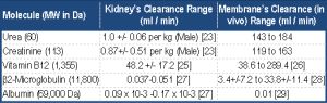 | Table 2- Clearance Range of Selected Molecules in the Kidney vs. Hemodialysis Membranes |
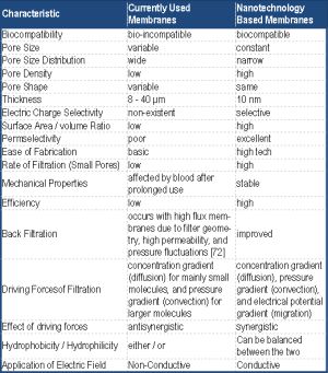 | Table 3- Difference Between Currently Available Membranes and Future Nanotechnology Based Membranes |