


ISSN : 0976-8920
EISSN : 0976-8939



PANDURANG C.1, DEVINDRA S.2*, SRINATH R.3
1Food Chemistry Division, National Institute of Nutrition (ICMR), Hyderabad-500007, AP, India.
2Food Chemistry Division, National Institute of Nutrition (ICMR), Hyderabad-500007, AP, India.
3Food Chemistry Division, National Institute of Nutrition (ICMR), Hyderabad-500007, AP, India.
* Corresponding Author : devindravs@indiatimes.com
Received : 27-09-2012 Accepted : 22-11-2012 Published : 06-12-2012
Volume : 3 Issue : 3 Pages : 87 - 89
J Crop Sci 3.3 (2012):87-89
DOI : http://dx.doi.org/10.9735/0976-8920.3.3.87-89
Conflict of Interest : None declared
Callus was inducted from various explants viz., cotyledon, hypocotyls and leaf explants of in vitro grown plantlets in sunflower. Two auxins i.e. 2, 4-D and NAA were used for callus induction in the range of 1-3 at 2 and 3mg/1 responded well for callus induction and growth NAA did not favored much callus induction at any of the concentration used. Supplementing BAP and Knowledge along with auxins further enhanced the growth of callus Knowledge being superior over BAP. Regeneration was obtained from cotyledon derived callus on MS + 0.5mg/1 IAA. The plantlets were rooted on MS medium supplemented with 2mg/1 NAA.
Helianthus annus, callus, multiple shoots.
Sunflower (Helianthus annus L) belongs to family astaraceae. The plants are annual herbs growing to a height of 4 to 10 ft. It is the third important major edible oil seed crop in the world after soybean and groundnut. The major bottleneck in quality improvement and production is the susceptibility of this plant to fungal disease like Alternaria blight, rust disease, downy mildew and root/color rot. These disease causes to yield loss up to 80%. Among options open to plant breeder to widen the genetic base are exploitation of alien variation. It is highly pertinent to devise and adopt new and conventional means to complement the traditional methods of breeding for disease resistant sources available [1] , other option is selection of disease resistant cell lines and regeneration of plantlets from the resistant cell lines. However, information on the cultural conditions favoring callus induction and subsequent regeneration is a prerequisite for the utilization of this technique.
Sunflower regenerability by organogenesis is highly variable and depends upon genotype, specific media components and nature of explant [2] . Further it is reported in this species that plant regeneration parameters have been shown to be under quantitative genetic control [3] . The present work is taken up with a view of inducting callus from various explants and subsequent regeneration. This protocol can be used for selection of cell lines and regenerating plantlets resistant to biotic and abiotic stress.
Seeds of sunflower var APHS-11 were obtained from agricultural research station, Gulbarga. The seeds were treated with 5% Bavistin and surface sterilized with 0.1% mercury chloride (HgCl2) for 3 min. and washed thrice with double distilled water inside the laminar airflow chamber to remove the traces of mercury chloride (HgCl2). The seeds were germinated in distilled water in culture tubes over a filter paper bridge. The aseptically germinated seedlings were used for the further studies. Various explants such as cotyledon hypocotyls and leaf were inoculated on Murashige and Skoog’s, [4] medium fortified with different concentrations of auxins (2, 4-D and NAA) alone or in combination with cytokinins (BAP and Knowledge) pH of the medium was adjusted to 5.8 then solidified with agar (0.8%) and autoclaved at 121°C for 15 minutes. The cultures were incubated at 27±2°C with 16 hrs. photoperiod.
Cotyledon, hypocotyl and leaf explants were inoculated on MS medium fortified with different concentrations of 2, 4-D and NAA (1-3mg/1). Callus initiation occurred with in 3 to 4 days from cotyledon and hypocotyls explants, while initiations of callus were observed in leaf explants only after 10 days. The frequency of callus induction was 100% in cotyledonary explant followed leaf (80.40%) and hypocotyl explant (70%) [Table-1] .
To investigate the effect of different auxins alone or in combination on the growth of callus, approximately 150±10mg of callus was inoculated on MS medium fortified with different concentrations (1-3mg/1) of auxin 2, 4-D and NAA. 2, 4-D AT 1, 3 & 3mg/1 supported growth of calli, however at mg/1, 2, 4-D fresh and try weight of callus decreased when compared to callus grown at 2mg/1 [Table-2] . However Lupi [5] reports that in hypocotyls explant induced callusing on BAP alone. Kiranmal and Prathibadevi [6] have reported that auxins did not favored callusing from cotyledons. However they may be due to formation on MS + 2mg/1 BAP. This differentiate behavior of explants may be due to different cultivars used, thus indicating callusing response and response to growth regulators is highly depended on genotype.
NAA at all the concentration evoked little growth response and only roots formed from the explants (Data not shown).
There are reports that supplementing small amounts (0.25 to 1mg/1) of cytokinins enhances the growth of callus in sunflower [6,7] and in several other species such as Niger [8] Cicer [9] ) Cajanus cajan [10] Vigna Radiata [11] ). Since 2, 4-D at 2mg/1 gave good results, it was felt to supplement auxins such as BAP and Knowledge along with 2, 4-D to study the interaction of 2, 4-D and auxins on the growth of callus. From the results presented in [Fig-1] & [Fig-2] it is clear that BAP (1mg/1) and Knowledge (0.5mg/1) supported further growth of callus when supplemented along with 2, 4-D and Knowledge proved superior over BAP.
For regeneration studies 200±20mg of callus was transferred to MS medium supplemented with 0.5 to 3mg/1 BAP. On medium supplemented with lower concentration (0.5 to 1mg/1) callus turned green and was compact initiation of shoot buds was observed after 10 days of transfer [Fig-3] . Ultimately the shoot buds elongated on medium supplemented with 0.5mg/1 BAP + 3mg/1 IAA. Small pieces of calli with shoot buds when transferred to MS medium supplemented with 0.5mg/1 BAP + 3mg/1 IAA elongation of shoots occurred [Fig-4] .
BAP induced organogenesis various explants has been reported earlier [12-14] from cotyledonary explant and Lupi [5] from cotyledonary callus.
For inducing roots 3-4 cm long plantlets were inoculated on MS medium supplemented with various concentrations of (1-3mg/-1) NAA and IBA. The data is presented in [Table-2] . Among the two auxins NAA at 2mg/1 induced maximum roots [Fig-5] & [Fig-6] . The rooted plantlets were removed and adhering agar with cleaned to transferred to plastic cups with 1:1 sterilized sand and soil mixture for hardening [Fig-6] . NAA and IBA induced rooting in the species has been reported by earlier workers [15-18] .
[1] Bajaj Y.P.S., Kumar P., Labaua K.S., Singh M.M. (1981) Indian Journal Experimental Biology, 19, 1026-1029.
» CrossRef » Google Scholar » PubMed » DOAJ » CAS » Scopus
[2] Berrior E.F., Gentzbittel L., Seriyes H., Alibert G., Sarrafi A. (1999a) Plant Cell, Tissue Organ Culture, 59, 65-69.
» CrossRef » Google Scholar » PubMed » DOAJ » CAS » Scopus
[3] Berrior E.F., Gentzbittel L., Seriyes H., Alibert G., Grievan Y., Berville A., Sarrafi A. (1999b) Plant Breeding, 118, 359-361.
» CrossRef » Google Scholar » PubMed » DOAJ » CAS » Scopus
[4] Murashgi T., Skoog’s F. (1962) Physiology Plantarum, 15, 473-497.
» CrossRef » Google Scholar » PubMed » DOAJ » CAS » Scopus
[5] Lupi M.C., Bennici A., Louci F., Gennai D. (1987) Plant Cell Tissue and Organ Culture, 11(1), 47-55.
» CrossRef » Google Scholar » PubMed » DOAJ » CAS » Scopus
[6] Kiranmai V., Prathiba Devi. (1995b) Advance Plant Science, 3(2), 338-341.
» CrossRef » Google Scholar » PubMed » DOAJ » CAS » Scopus
[7] Dhaka N., Kothari S. (2002) Plant Cell Report, 21, 29-34.
» CrossRef » Google Scholar » PubMed » DOAJ » CAS » Scopus
[8] Sarvesh A. (1990) Ph.D. thesis submitted Osmania University, Hyderabad, India.
» CrossRef » Google Scholar » PubMed » DOAJ » CAS » Scopus
[9] Rao Srinath., Naidu M. (1989) Proceedings of Indian Academic Science (Plant Science), 99 (6), 523-527.
» CrossRef » Google Scholar » PubMed » DOAJ » CAS » Scopus
[10] Srinath Rao., Basavaraj K. (1990) Indian Journal of Botany, 13, 192-196.
» CrossRef » Google Scholar » PubMed » DOAJ » CAS » Scopus
[11] Srinathrao., Prabhavati Patil., Kaviraj C.P. (2005) Indian Journal of Biotechnology, 4, 556-560.
» CrossRef » Google Scholar » PubMed » DOAJ » CAS » Scopus
[12] Deglene L., Lesignes P., Alibert G., Sarrafi A. (1997) Plant Cell Tissue and Organ Culture, 48, 127-130.
» CrossRef » Google Scholar » PubMed » DOAJ » CAS » Scopus
[13] Nataraj K., Ganapathi T.R. (1989) Indian Journal of Experimental Biology, 27, 777-779.
» CrossRef » Google Scholar » PubMed » DOAJ » CAS » Scopus
[14] Ceriani M.F., Hopp H.E., Hahne G., Escandon A.S. (1992) Plant Cell Physiology, 33(2), 157-164.
» CrossRef » Google Scholar » PubMed » DOAJ » CAS » Scopus
[15] Epinasse A., Lay C., Volin J. (1989) Plant Cell Tissue Organ Culture, 17(13), 171-181.
» CrossRef » Google Scholar » PubMed » DOAJ » CAS » Scopus
[16] Witizens B., Scowcroft W.R., Dowens R.W., Larkin P.J. (1988) Plant Cell Tissue Organ Culture, 13, 61-76.
» CrossRef » Google Scholar » PubMed » DOAJ » CAS » Scopus
[17] Pandurang C. (2000) Ph.D. thesis submitted to Gulbarga University, Gulbarga, India.
» CrossRef » Google Scholar » PubMed » DOAJ » CAS » Scopus
[18] Ramagopal Rao S. (2002) Ph.D. thesis submitted Gulbarga University, Gulbarga, India.
» CrossRef » Google Scholar » PubMed » DOAJ » CAS » Scopus
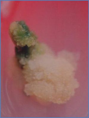 | Fig. 1- Effect of BAP on the growth of callus when supplemented along with 2, 4-D |
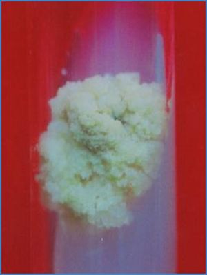 | Fig. 2- Knowledge proved superior over BAP. |
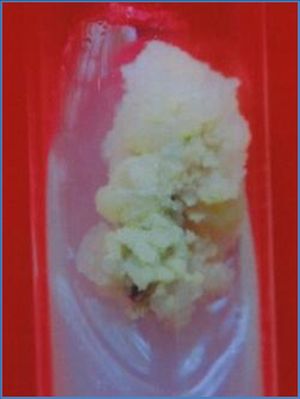 | Fig. 3- Callus turned green and was compact initiation of shoot buds was observed after 10 days of transfer. |
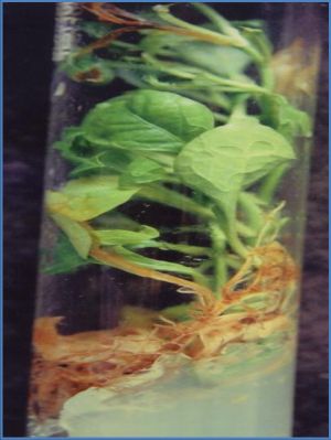 | Fig. 4- Small pieces of calli with shoot buds when transferred to MS medium supplemented with 0.5mg/1 BAP + 3 mg/1 IAA elongation of shoots occurred. |
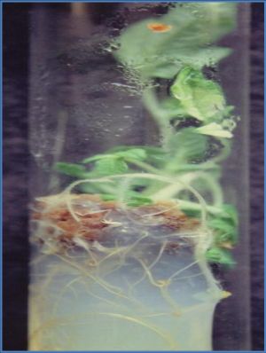 | Fig. 5- Among the two auxins NAA at 2 mg/1 induced maximum roots |
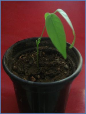 | Fig. 6- Plantlets of Sunflower. |
 | Table 1- Number of days required for callusing and frequency of callus induction of different explants in sunflower on MS medium supplemented with 2mg/1m 2, 4-D |
 | Table 2- Effect of 2, 4-D on the growth of callus derived from cotyledonary explant in sunflower |