


ISSN : 0976-8726
EISSN : 0976-8734



DEEPAK N.1*, PANCHASHEELAN2, ANILKUMAR T.3, RAVIKUMAR4, JAYAKUMAR5
1Department of General Medicine, ESIC-MC PGIMSR, Rajajinagar, Bangalore-560010, Karnataka, India.
2Department of General Medicine, ESIC-MC PGIMSR, Rajajinagar, Bangalore-560010, Karnataka, India.
3Department of General Medicine, ESIC-MC PGIMSR, Rajajinagar, Bangalore-560010, Karnataka, India.
4Department of General Medicine, ESIC-MC PGIMSR, Rajajinagar, Bangalore-560010, Karnataka, India.
5Department of General Medicine, ESIC-MC PGIMSR, Rajajinagar, Bangalore-560010, Karnataka, India.
* Corresponding Author : deepak.n.cool@gmail.com
Received : 16-10-2012 Accepted : 20-11-2012 Published : 20-12-2012
Volume : 3 Issue : 1 Pages : 34 - 36
Med Case Rep 3.1 (2012):34-36
DOI : http://dx.doi.org/10.9735/0976-8726.3.1.34-36
Conflict of Interest : None declared
Strongyloidiasis or threadworm infection is endemic in tropical and subtropical climate caused by the parasitic nematode Strongyloides stercoralis. Infection may be asymptomatic producing minimal symptoms or can be life threatening producing disseminated infection in immunocompromised individuals. A 55 year old male presented with pain abdomen, vomiting, fever of 1 year duration. He had significant weight loss of 24 kgs(from 58 to 34 kgs) in 6 months. Upper GI endoscopy was done to rule out malabsorption disorder and duodenal biopsy was taken and sent for histopathology which showed strongyloides stercoralis worms. Stool samples were sent to search for strongyloides larvae. Stool examination was positive for rhabditiform larvae of strongyloides stercoralis confirming diagnosis. Patient was started on Ivermectin 200mcg/kg. Repeat stool examinations done after 1 week of starting therapy were negative for strongyloides larvae.This case report highlights the importance of strongyloidiasis as a cause of malabsorption and its diagnosis by duodenal biopsy.
Strongyloidiasis, Malabsorption, Hyperinfection, Disseminated infection, Cachexia.
Strongyloidiasis or threadworm infection is endemic in tropical and subtropical climate caused by the parasitic nematode Strongyloides stercoralis. Infection may be asymptomatic producing minimal symptoms or can be life threatening producing disseminated infection in immunocompromised individuals. Its characteristic ability is in producing hyperinfection syndrome by autoinfection thereby persisting in the host for decades. The second species of this parasite S. fuelleborni produces life threatening swollen belly syndrome in infants with features of generalised edema and respiratory distress. It infects humans in areas of Africa and papua new guinea. Here we reort a case of Strongyloidis stercoralis leading to severe cachexia [1-3] .
A 55 year old male presented with pain abdomen, vomiting, fever of 1 year duration. He had significant weight loss of 24kgs (from 58 to 34kgs) in 6 months. His general physical examination revealed signs of severe malnutrition and multiple vitamin deficiencies. Complete blood counts were normal except for mild anemia(Hb-9.2g%). Blood urea and serum creatinine were normal. He had severe hypoalbuminemia (1.2g%). Ultrasound abdomen showed mild hepatomegaly. His initial stool examination was negative for ova, cysts. Thyroid function test was normal. HIV serology was negative. He was a known diabetic from 1 year on irregular treatment. Upper GI endoscopy was done to rule out malabsorption disorder and duodenal biopsy was taken and sent for histopathology which showed strongyloides stercoralis worms. Stool samples were sent again to search for strongyloides larvae. Stool examination was positive for rhabditiform larvae of strongyloides stercoralis confirming diagnosis. Patient developed pyothorax which was treated with higher antibiotics and intercostal drainage. Patient was started on Ivermectin 200mcg/kg. Repeat stool examinations done after 1 week of starting therapy were negative for strongyloides larvae.
[Fig-1] describes life cycle of Strongyloides stercoralis.
The nematode Strongyloides reproduces by parthenogenesis and releases eggs into the intestinal lumen from which rhabditiform larvae emerge and excreted in feces. Rhabditiform larvae either develop into free-living adult male and female worms or infectious filariform larvae. Infectious filariform larvae penetrate the skin of humans and enter the venous circulation and then pass to the lungs, which are swallowed and pass through the stomach, and adult female worms develop in the small intestine.
Small numbers of rhabditiform larvae develop into filariform larvae within the intestines and reenters through the colonic mucosa or the perianal skin producing autoinfection. As a result worms increase in large numbers without exogenous reinfection and persist in the same host for longer duration [2,3] .
• Corticosteroids and other immunosuppressive agents(chemotherapeutic agents, tacrolimus, tumor necrosis factor alfa inhibitors).
• Corticosteroids and other immunosuppressive agents (chemotherapeutic agents, tacrolimus, tumor necrosis factor alfa inhibitors) [3,4] .
• Human T-cell leukemia virus type 1 (HTLV-1) infection [3,4] Human immunodeficiency virus (HIV) infection [4] .
• Hypogammaglobulinemia.
• Hematologic malignancies (lymphoma, leukemia) [3] .
• Organ transplantation [3,4] .
• Malabsorption states and malnutrition.
• Chronic renal failure and end-stage renal disease [3] .
• Diabetes mellitus.
• Advanced age.
• Collagen-vascular disease.
• Chronic alcohol consumption.
Most of the infections are asymptomatic in immunocompetent persons or may be associated with mild gastrointestinal manifestations such as diarrhea, abdominal pain or bloating. Rarely gastrointestinal bleeding, small bowel obstruction, paralytic ileus may result.
Migration of larvae through the skin may cause Larva currens, allergic reaction characterised by serpiginous erythematous maculopapular tracks and urticarial lesions on the skin of the buttocks, thighs, and lower abdominal wall. Migration of larvae through lungs may lead to condition resembling Loeffler’s syndrome [1-3] .
Immunosuppression of any cause may lead to hyperinfection and disseminated syndrome [3,4] .
It occurs due to accelerated production of filariform larvae in immunosuppressed state. With hyperinfection, increased numbers of larvae are found in the intestines and lungs and with disseminated strongyloidiasis, larvae are also found in the central nervous system (CNS), kidneys, liver, and almost any other organ. Eosinophilia is usually absent [4] .
The Gastro intestinal manifestations include abdominal pain, watery diarrhea, weight loss, nausea, vomiting and occasionally gastrointestinal bleeding, peritonitis. The symptoms may resemble that of ulcerative colitis. Edema of the bowel may lead to Subacute intestinal obstruction [1,3] .
The extraintestinal manifestations include pneumonitis with cough, hemoptysis and respiratory failure with diffuse bilateral infiltrates on the chest x ray [1,4] eosinophilic pleural effusions and eosinophilic granulomatous enterocolitis may be seen in strongyloidiasis.
Bacterial sepsis, meningitis, and pneumonia are frequent in disseminated infections.
The cutaneous manifestations that could occur from dissemination include widespread petechiae and purpura.
Gram negative bacteremia mainly from pathogens such as Streptococcus bovis, Escherichia coli, Streptococcus fecalis, Klebsiella pneumoniae or Enterobacter species can lead to fatal complications as they become blood borne when the larvae penetrate the intestine [1,3] .
Eosinophilia may be the first clue for the diagnosis of intestinal strongyloidiasis but it is mild and nonspecific. Eosinophilia is usually absent in disseminated infection [3-6] .
The serological methods determine the presence of strongyloid antibody in the serum of the human hosts. The antibody could be determined by the following methods.
• Enzyme linked immunosorbent assay (ELISA).
• Gelatin particle indirect agglutination (GPIA).
• Western blot analysis (WBA).
Microscopic examination of stool specimen is done for strongyloid larvae [Fig-2] . This can be in the following way:
• Simple direct smear with sensitivity of 0-52%.
• Formalin-ether concentration method with a sensitivity of 13-55%.
• Harada-Mori filter paper culture method with almost equal sensitivity of 13-55%.
• Agar plate culture technique with higher sensitivity of 78-100%.
Examination of duodenal aspirate is found to be highly sensitive for diagnosis of strongyloidiasis especially in immunocompromised children. Parasites are found only in duodenal aspirate and not in faeces in some cases. However it is invasive and not routinely performed [1,6] .
Histopathological examination of duodenal biopsy specimen may identify strongyloides worms or larvae embedded in the mucosa as seen in our case [Fig-3] .
• Ivermectin at a dose of 200mcg/kg for 2 days is effective for most uncomplicated infections.
• For hyperinfection and disseminated syndrome, treatment is continued for minimum of 7 days until larvae are eradicated from stool, sputum and urine.
• Thiabendazole and albendazole are alternatives.
• Antibiotic Therapy directed against enteric pathogens if bacteremia is present and Supportive Care including intravenous fluids may be required [1-3] .
Inspite of recent advances, strongyloidiasis has still been an elusive disease. Strongyloidiasis needs special attention by the physician especially in malnourished, debilitated patients. strongyloidiasis as a cause of malabsorption must be kept in mind while evaluating chronic debilitated conditions.
[1] Mandell G.L., Bennett J.E., Dolin R. Mandell, Douglas and Bennetts (2009) Principals and Practice of Infectious Diseases. 7th ed.
» CrossRef » Google Scholar » PubMed » DOAJ » CAS » Scopus
[2] Kliegman R.M., Stanton B.F., Gemelll J.W., Schor N.F., Behrman R.E. (2011) Textbook of Paediatrics., 19th ed.
» CrossRef » Google Scholar » PubMed » DOAJ » CAS » Scopus
[3] Goldman L., Schafer A.I. (2011) Goldman’s Cecil Medicine, 24th ed.
» CrossRef » Google Scholar » PubMed » DOAJ » CAS » Scopus
[4] Vadlamudi R.S., Chi D.S., Krishnaswamy G. (2006) Clinical and Molecular Allergy.
» CrossRef » Google Scholar » PubMed » DOAJ » CAS » Scopus
[5] Concha R., Harrington W.J., Rogers A.I. (2005) J. Clin. Gastroenterol., 39, 203-211.
» CrossRef » Google Scholar » PubMed » DOAJ » CAS » Scopus
[6] Siddiqui A.A., Berk S.L. (2001) Clin. Infect Dis., 33, 1040-1047.
» CrossRef » Google Scholar » PubMed » DOAJ » CAS » Scopus
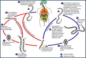 | Fig. 1- Life cycle of Strongyloides stercoralis |
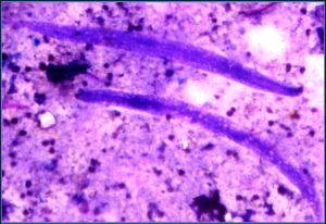 | Fig. 2- Stool examination showing rhabditiform larvae |
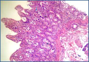 | Fig. 3- Histopathological examination of duodenal biopsy specimen (Hematoxylin &Eosin stain) Black arrow- rhabditiform larvae within egg Blue arrow- adult worms |