


ISSN : 0976-996X
EISSN : 0976-9978



SILVA G.F.R.1, BRANDÃO L.N.S.2, PAULA D.A.J.3, PESCADOR C.A.P.4, CHITARRA C.S.5, CARVALHO R.C.T.6, NAKAZATO L.7, DUTRA V.8*
1Microbiology Veterinary Laboratory, Federal University of Mato Grosso (UFMT), Brazil.
2Microbiology Veterinary Laboratory, Federal University of Mato Grosso (UFMT), Brazil.
3Microbiology Veterinary Laboratory, Federal University of Mato Grosso (UFMT), Brazil.
4Pathology Veterinary Laboratory, Federal University of Mato Grosso (UFMT), Brazil.
5Microbiology Veterinary Laboratory, Federal University of Mato Grosso (UFMT), Brazil.
6Molecular Biology Veterinary Laboratory, Federal University of Mato Grosso (UFMT), Brazil.
7Molecular Biology Veterinary Laboratory, Federal University of Mato Grosso (UFMT), Brazil.
8Microbiology Veterinary Laboratory, Federal University of Mato Grosso (UFMT), Brazil.
* Corresponding Author : valdutra@ufmt.br
Received : 16-08-2012 Accepted : 21-09-2012 Published : 24-10-2012
Volume : 3 Issue : 1 Pages : 55 - 59
Vet Sci Res 3.1 (2012):55-59
DOI : http://dx.doi.org/10.9735/0976-996X.3.1.55-59
Conflict of Interest : None declared
The aim of this study was to characterize Brazilian isolates of P. multocida from pig lungs based on Multilocucs sequence typing (MLST) and virulence factors. We analyze 27 isolates of P. multocida from lungs with pneumonia and 2 isolates from lungs without lesions. PCR was used to confirm P. multocida by kmtI gene and identify the presence of virulence factors genes toxA, pfhA and tbpA. Almost all samples belong to serotype A (27/29) were from pneumonic lesions and only two of serotype D (2/29) were from healthy lung. No samples amplified gene toxA. Sixteen samples (55,1%) amplified the tbpA and ten (34,48%) the pfhA. Lincosamid and aminoglycosides resistance were observed in 80% and 52% of isolates, respectively. Upon MLST analysis, 13 genotypes were identified, nine isolates have ST that was not previous reported. Occurrence of Clonal Complex (CC) in pneumonic lungs was associated to CC74 that have seventeen isolates followed by CC13 with four isolates. No pneumonic lungs have CC50 detected with two isolates. Six isolates (ST 190, 193, 194, 198, 197) have no CC identity to previous complex but concatenate phylogenetic analysis showed a closely relationship with CC74 and CC13. Phylogenetic analysis shows that all isolates sequence type in this study has a linkage disequilibrium (IsA=0,40; P=0,000) and probably this swine isolates have a clonal distribution. P. multocida isolated from swine pneumonic lungs at Central Western Brazil region have serotype A, great variability with two main CC (CC74 and CC13) complexes with a high number of isotates positive to tbpA virulence factor. This data could help to uncover P. multocida genetic diversity associated to swine pneumonia.
Pasteurellosis, virulence, toxA, pfhA, tbpA, MLST.
Pasteurella multocida is part of the microbiota of the respiratory tract of pigs, although some strains are associated with progressive atrophic rhinitis and others with pleurisy and pneumonia. These diseases are highly contagious and responsible for significant losses in modern swine production [1-3] . Currently there are five capsular serotypes (A, B, D, E and F) of P. multocida [1] and several authors have studied the presence of virulence genes involved in the pathogenesis of pneumonia caused by P. multocida with toxA, pfhA, tbpA, ompH, ptfA, hgbA, pls and Nanh are the most studied [3-6] . Some virulence factors are associated with pathogenicity, including the dermonecrotic toxin gene (toxA), filamentous hemagglutinin gene (pfhA) and transferrin binding protein gene (tbpA) [16] .
Recently, Multiloccus Sequencing Typing (MLST) technique were developed to characterized P. multocida from avian cholera [7] and then applied to bovine and swine pneumonia isolates [8] . This has advantage to be portable and results comparable between laboratories. Since no data is available based on this technique, the objective of this study was to characterize isolates of P. multocida from lungs of pigs with pneumonia and demonstrate population structure associated of P. multocida associated to pneumonia in Central-western of Brazil.
One hundred samples were collected from lungs of pigs, forty with and sixty without macroscopic lesion of pneumonia, at slaughterhouse under Federal inspection in Mato Grosso state in the period December 2009 to March 2011 [Table-1] Five samples were collected from outbreaks of pneumonia occurred in the same period [Table-1] The macroscopic changes of the lungs for inclusion in the study were: hepatization, fibrin deposition, pleurisy or adherence. Fragments were kept on ice and fixed in 10% formalin and sent to the laboratory for microbiological and histopathological processing.
The samples were plated on 5% sheep blood and MacConkey agar, incubated at 37°C for 48 hours. The samples that showed the coccobacilli morphology and negative on MacConkey agar cultivation were subjected to biochemical tests of catalase, oxidase, urease, triple sugar iron (TSI), glucose, sucrose, lactose, maltose and mannitol. The isolates classified as P. multocida [9] were submitted to antibiogram test by disk diffusion method [10] . The antimicrobial agents tested included oxaciclin (1 mg), norfloxacin (10 mg), lincomycin (2 mg), tetracycline (30 mg), ampicillin (10 mg), colistin (10 mg), streptomycin (10 mg), ofloxacin (5 mg), enrofloxacin (5 mg), doxycycline (30 mg), amoxicillin (10 mg), ceftiofur (30 mg) and penicillin (10 IU).
P. multocida classified by biochemical tests were grown in a medium (BHI) for 24 hours at 37°C with stirring and subjected to extraction of genomic DNA [11] . Isolates of P. multocida were confirmed by kmtI gene PCR (specific to P. multocida) and capsular typing was determined with specific genes, hyaD-hyaC, bcbD, dcbF, ecbJ and fcbD to identify the serotypes A, B, D, E and F, respectively [12] . The presence of genes of virulence factors toxa, tbpA and pfhA were determined [5] . The amplification products were analyzed by electrophoresis on 2% agarose gel stained with ethidium bromide (10μg/mL) for 1 h at 125V per cm UV transilluminator and observed with marker ladder.
Lungs fragments were immersed in formalin 10% for 24 hours, processed for histopathology and stained with hematoxylin-eosin and observed in optical microscopic [13] .
Multilocus sequence typing scheme and protocol from P. multocida RIRDC MLST Databases were realized on all isolates [7] . Genetic analysis were performed on START2 software [14] and multiple sequence alignment were done with Muscle and Neigborn Join with 100 bootstrap were utilized to construct concatenated phylogenetic tree from sequence of seven genes of P. multocida RIRDC MLST Databases captured November 21, 2011.
In 27 (60%) pneumonic lungs, from nine herds, had growth of P. multocida, confirmed by kmtI gene PCR amplification. The hyaD-hyaC gene was amplified all samples classifying them as belonging to serotype A [Table-1] . Only two (3.3%) isolates were detected in healthy lung and they were serotype D. Histophatological findings were depicted in [Table-3] . Bronchopneumonia and interstitial pneumonia were detected at variable degree, distribution and edema in lung with pneumonia. No microscopic lesions were detected in lung whitout macroscopic lesion.
Analysis of virulence factors shows that at least one gene (tbpa, toxA e pfha) were detected in 25 isolates (92%). Virulence factor show eleven out of 27 (34.48%) isolates with pfha gene and sixteen (55.17%) with tbpa gene. All isolates were negative to toxA. The samples showed 80% resistance to lincosamids and 52% had partial sensitivity to aminoglycosides. To other antibiotics (quinolone, tetracycline, polypeptides and cephalosporin), sensitivity was high, 80 to 100% [Table-2] .
Under MLST analysis, in this study 13 genotypes were described from 29 P. multocida isolates. Four genotypes were previous described and nine were new and submitted to PUBMLST. Isolate M225 have new alleles to adk, est, gdh and pgi genes [Fig-1] . Comparison between isolates in this study and isolates from pig in P. multocida (RIDC) database shows a presence of linkage disequilibrium (IsA=0,40; P=0,000).
Clonal complex analysis with five of seven identical alleles genes show presence of CC 74, CC13 and five STs without previous described in RIDC database. Concatenate analysis with all ST of RIDC database show that this isolates are phylogenetic closely related to CC13 (ST197) and CC74 (ST190, ST193, ST194 and ST198) [Table-4] . At herd level, CC 74 was the most frequent occurring in five herds followed by CC13 in two herds. CC50 was detected in herd associated to lung with no macroscopic lesion.
P. multocida serotype A is responsible for pneumonia in pigs worldwide. In the UK, a total of 129 strains isolated from lungs of pigs with pneumonia, 106 (82.2%) belonged to serotype A [15] . In Germany of 155 samples of P. multocida isolated from swine pneumonia, 99 (63.9%) belonged to serotype A [4] . In South Korea, 30 samples of P. multocida in pigs with pneumonia, 17 (56.6%) belonged to serotype A and 13 (43.4%) belonged to serotype D [16] . Unlike in China of 233 samples from pigs with clinical signs of respiratory disease 92 (39.5%) belonged to serotype A and 128 (54.9%) to D [3] , but in China the samples analyzed were from throughout the respiratory tract different of studies where the samples came just lung, this may be the justification for the higher number of serotype D found in China.
In Brazil, Borowski S.M., et al [1] ; Moreno A.M., et al [17] ; Heres T.S. [18] also isolated the serotype A in most samples of lungs from pigs with pneumonia (95.5%, 89.4% and 93.91%, respectively). Analyzing only samples of the Mato Grosso and South Mato Grosso States, studied by Moreno A.M., et al [17] 100% were serotype A, a result similar to this study. P. multocida serotype D was the most isolated in pneumonia in Santa Catarina State [19] , however unlike this study chronic abscess lesions were the most found. toxA gene was not observed in this study. Studies conducted in the UK, Davies R.L., et al [15] identified this gene in 9% of samples. 6.3% Ewers C., et al [5] and Bethe A., et al [4] in Germany, in China this gene only was identified in samples of P. multocida serotype D [3] . In Brazil, this gene was not identified recently [18] , however a few years ago was identified in 9 (37.5%) [20] and one (1.72%) [17] samples from pigs with pneumonia.
toxA gene is responsible for transcription of the dermonecrotic toxin and although it is related to the virulence of strains of P. multocida, some authors identify the most commonly in cases of atrophic rhinitis or pneumonia caused by strains of P. multocida serotype D [3-5,20] . The absence or low presence of this gene in isolates of P. multocida serotype A of pig’s lungs with pneumonia in this and other studies [3,5,17] can be explained by the greater importance of toxA gene in the pathogenesis of atrophic rhinitis caused by serotype D of P. multocida [5] .
pfhA gene amplified in 10 (34,48%) of the isolates, higher than that found by others authors [4,5] . This gene had a higher presence from P. multocida isolated from diseased animals (23.3%) than in carriers (15.4%) [4] . Among diseased animals pfhA gene was more prevalent in animals with lower respiratory tract infections when compared with isolates from animals with upper respiratory tract infection [4] . In China Tang X., et al [3] identified the presence of this gene in 23 (25%) samples isolated from P. multocida serotype A, while only 4 (3.1%) samples belonging to serotype D amplified the gene pfhA. In addition, this aothors described a relationship between the presence of this gene with pneumonia of pigs and cattle. pfhA gene is responsible for the production of filamentous hemagglutinin, the protein is of the same class of Bordetella pertussis FHA protein and contributes to the colonization and adherence of the bacteria in the nasopharynx and trachea of ​​the affects animals [21] .
tbpA gene was amplified in 16 (55.17%) isolates from pneumonic lungs. This gene were not found in isolates of P. multocida pneumonia in pigs in others studies [3-5] , however the authors considered the possibility that this gene is involved in virulence of isolates of P. multocida outbreaks of hemorrhagic septicemia in ruminants and other animals [5] . The high occurrence of this gene in this study may reflect regional characteristic and explained by the even higher correlation with pneumonia in ruminants and with P. multocida serotype B, that was not identify in this study [10] . In India the presence of tbpA gene across species and among these pigs and identified as an important marker of pneumonia in epidemiological several species where it is present [22] .
Bethe A., et al [4] suggests that the presence or absence of some genes such as toxA, pfhA and tbpA may be associated with the virulence of strains of sick animals and can be utilized as an important epidemiological marker. According to the results obtained, it is suggested that the gene pfhA can be used as a marker for pneumonia in pigs, because of pneumonia among the samples this gene had a high occurrence. However pneumonia by P. multocida in pigs is a multifactorial disease, where several factors such as nutrition, stocking rate, climate and other environmental characteristics contribute to the maintenance of the disease in swine herd [23] . Other studies focused on protein expression are needed to explain the pathogenesis of pneumonia caused by P. multocida in pigs.
Several authors have shown that isolated P. multocida are resistant to lincosamides [1,24] , although they identified an increased resistance to multiple drugs than that observed in this study. In addition to resistance to lincosamides and β-lactam was also observed resistance to quinolones [20,24] and tetracycline [20] .
This resistance can be justified by the indiscriminate use of antimicrobials in pig farming state, both for the treatment of pathologies such as the use of growth promoters. in a retrospective study from 2003 to 2007 showed that the resistance of strains of P. multocida in China increased over the years, from 47% in 2003 to 97% in 2007 to more than 5 antibiotics [3] , which shows concern for the future treatment of pneumonia in the state of Mato Grosso and Mato Grosso do Sul.
The microscopic lesions found were subacute suppurative bronchopneumonia (50%) and interstitial pneumonia (50%) [Table-3] . Others authors in Brazil also observed the presence of suppurative bronchopneumonia or hemorrhagic necrosis and subacute in pigs free of respiratory pathogens that after inoculation of P. multocida serotype A reproduced the disease with the same serotype and microscopic lesions similar to this study [25] . In Denmark, 28 cases of acute or subacute suppurative bronchopneumonia with isolation of P. multocida were observed [25] , results similar to ours, however, in these cases there was concomitant identification of Mycoplasma hyopneumoniae, unlike this study where only five samples were positive for M. hyopneumoniae and one for Haemophillus parasuis by PCR, according to Cai, et al [26] and Oliveira, et al [28] respectively.
Under MLST analysis, in a study in Denmark, 25 isolates of different PFGE (pulsed-field gel electrophoresis) profile were analyzed by MLST with identification of seven sequence types (ST 13, 50, 74, 148, 149 and 150) that were part of three clonal complex (CC 13, 50 and 74) when consider five out of sevem similarity alleles. Isolates in this study shows a more genetic diversity with 13 STs in the 28 isolates and with three previous described CC (C13, 50 and 74) and more five new CC. CC13 and CC74 were described in pig in other countries isolated mainly from pneumonic lesions. ST 27 is classified as CC 74, but was not described previously in pig. ST191, 193, 194, 197 and 198 has no CC associated.
Phylogenitcs analys shows ST195 (CC50) was described in both in pneumonia [28] and atrophic rhinitis but this isolated were only detected in healthy pig lungs (pubmlst). In this database the occurrence of isolates without pneumonia is greather when compared to others CC 74 and CC13.
P. multocida isolated in Mato Grosso and Mato Grosso do Sul belong to serotype A and among them was the identification of genes pfhA (48%) and tbpA (4%), which has so far not been reported in Brazil. The samples were resistant to lincosamids and sensitive to quinolonas, β-lactams, tetracyclines, aminoglycosides, polypeptides and cephalosporin. Genetic diversity were common with 13 genotypes mainly from clonal complex CC74 and CC13.
To CAPES, FAPEMAT and CNPq for financial support.
[1] Borowski S.M., Ikuta N., Lunge V., Fonseca A., Marques E., Cardoso M. (2002) Pesquisa Veterinária Brasileira, 22(3), 97-103.
» CrossRef » Google Scholar » PubMed » DOAJ » CAS » Scopus
[2] Pijoan C. (2006) IWOA: Blackwell Publishing, 719-726.
» CrossRef » Google Scholar » PubMed » DOAJ » CAS » Scopus
[3] Tang X., Hao Z., Hu J., Wu B., Cai X., He Q., Chen H. (2009) J. Clin. Microbiol., 47(4), 951-958.
» CrossRef » Google Scholar » PubMed » DOAJ » CAS » Scopus
[4] Bethe A., Wieler L.H., Selbitz H.J., Ewers C. (2009) Vet. Microbiol., 139(1-2), 97-105.
» CrossRef » Google Scholar » PubMed » DOAJ » CAS » Scopus
[5] Ewers C., Lübke-Becker A., Bethe A., Kiebling S., Filter M. Wieler L.H. (2006) Vet. Microbiol., 114(3-4), 304-317.
» CrossRef » Google Scholar » PubMed » DOAJ » CAS » Scopus
[6] Harper M., Boyce J.D., Adler B. (2006) FEMS Microbiol. Lett., 265(1), 1-10.
» CrossRef » Google Scholar » PubMed » DOAJ » CAS » Scopus
[7] Subaaharam S., Blackall L.L., Blackall P.J. (2010) Vet. Microbiol., 141(3-4), 354-361.
» CrossRef » Google Scholar » PubMed » DOAJ » CAS » Scopus
[8] Hotchkiss E.J., Hodgson J.C., Lainson F.A., Zadoks R. (2011) BMC Microbiology, 11(1150), 2-8.
» CrossRef » Google Scholar » PubMed » DOAJ » CAS » Scopus
[9] Quinn P.J., Carter M.E., Markey B., Carter G.R. (1994) London: Wolfe.
» CrossRef » Google Scholar » PubMed » DOAJ » CAS » Scopus
[10] Bauer A.W. and Kirby E.M. (1966) Am. J. Clin. Pathol., 45(4), 493-496.
» CrossRef » Google Scholar » PubMed » DOAJ » CAS » Scopus
[11] Sambrook J. and Russel D.W. (2004) New York: Cold Spring Harbor Laboratory Press.
» CrossRef » Google Scholar » PubMed » DOAJ » CAS » Scopus
[12] Townsend M., Boyce J.D., Chung J.Y., Frost A.J., Adler B. (2001) J. Clin. Microbiol., 39(3), 924-929.
» CrossRef » Google Scholar » PubMed » DOAJ » CAS » Scopus
[13] Allen T.C. (1992) Washington: American Registry of Pathology, 53-58.
» CrossRef » Google Scholar » PubMed » DOAJ » CAS » Scopus
[14] Jolley K.A., Feil E.J., Chan M.S., Maiden M.C. (2001) Bioinformatics, 17(12), 1230-1231.
» CrossRef » Google Scholar » PubMed » DOAJ » CAS » Scopus
[15] Davies R.L., Maccorquodale R., Baillie S., Caffrey B. (2003) J. Med. Microbiol., 52, 59-67.
» CrossRef » Google Scholar » PubMed » DOAJ » CAS » Scopus
[16] Anh K.K., Lee Y.H., Ha Y.l., Kim D., Chae S., Kim C.H., Lee J.H., Kim S.H., Chae C. (2008) J. Comp. Pathol., 139(1), 51-53.
» CrossRef » Google Scholar » PubMed » DOAJ » CAS » Scopus
[17] Moreno A.M., Baccaro M.R., Ferreira A.J., Pestana de Castro A.F. (2003) J. Clin. Microbiol., 41(4), 1743-1746.
» CrossRef » Google Scholar » PubMed » DOAJ » CAS » Scopus
[18] Heres T.S. (2009) Caracterização de Amostras de Pasteurella Multocida Isoladas de lesões Pneumônicas Associadas ou não com Circovirose em suÃnos. Dissertação, Universidade Federal do Rio Grande do Sul.
» CrossRef » Google Scholar » PubMed » DOAJ » CAS » Scopus
[19] Mores M.A.S. (2006) Anatomopatologia e Microbiologia de lesões Pulmonares Responsáveis por Condenação de Carcaças em SuÃnos. Dissertação, Universidade Federal do Paraná.
» CrossRef » Google Scholar » PubMed » DOAJ » CAS » Scopus
[20] Borowski S.M., Silva S.C., Schrank I., Cardoso M. (2001) Arquivos da Faculdade de Veterinária, 29, 79-85.
» CrossRef » Google Scholar » PubMed » DOAJ » CAS » Scopus
[21] Fuller T.E., Kennedy M.J., Lowery D.E. (2000) Microbial Pathog., 29, 25-38.
» CrossRef » Google Scholar » PubMed » DOAJ » CAS » Scopus
[22] Shivachandra S.B., Kumar A.A., Amaranath J., Joseph S., Srivastava S.K., Chaudhuri P. (2005) Vet. Res. Commun., 29(6), 537-542.
» CrossRef » Google Scholar » PubMed » DOAJ » CAS » Scopus
[23] Dalla Costa O.A., Morés N., Sobestiansky J., Barione Júnior W., Piffer I.A., Paiva D.P., Amaral A.L., Guzzo R., Lima G.J.M.M., Perdomo C.C. (2000) Embrapa SuÃnos e Aves, 267, 1-5.
» CrossRef » Google Scholar » PubMed » DOAJ » CAS » Scopus
[24] Shin S.J., Kang S.G., Nabin R., Kang M.L., Yoo H.S. (2005) Vet. Microbiol., 106(1-2), 73-77.
» CrossRef » Google Scholar » PubMed » DOAJ » CAS » Scopus
[25] Kich J.D., Mores N., Triques N.J., Nogueira M.G., Locatelli C., Klein C.L., FelÃcio R.P. (2007) Embrapa SuÃnos e Aves, 469, 1-7.
» CrossRef » Google Scholar » PubMed » DOAJ » CAS » Scopus
[26] Cai H.Y., Dreumel T.V., Mcewen B., Hornby G., Bell-Rogers P., Mcraild P., Josephon G., Maxie G. (2007) J. Vet. Diagn. Invest., 19(1), 91-95.
» CrossRef » Google Scholar » PubMed » DOAJ » CAS » Scopus
[27] Oliveira S., Galina L., Pijoan C. (2000) J. Vet. Diagn. Invest., 13(6), 495-501.
» CrossRef » Google Scholar » PubMed » DOAJ » CAS » Scopus
[28] Pors S.E., Hansen M.S., Christensen H., Jensen H.E., Petersen A., Bisgaard M. (2011) Vet. Microbiol., 150(3-4), 354-361.
» CrossRef » Google Scholar » PubMed » DOAJ » CAS » Scopus
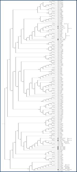 | Fig. 1- Concatenate sequence analysis tree was constructed using muscle alignment and Neighbor Joining (100 replicates) of seven genes of two hundred two sequence of sequencing typing (ST1 to ST202) from RIDC database. CC: clonal complex •; isolated with pneumonia isolated from lung without pneumonia. |
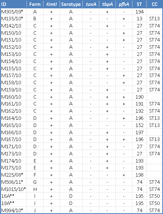 | Table 1 - Characteristic of 29 isolates of P. multocida from swine lungs from Mato Grosso and Mato Grosso do Sul States, during 2008 to 2011. *Outbreaks cases. |
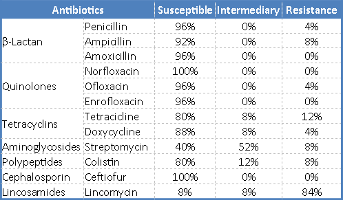 | Table 2- Profile of antimicrobial susceptibility of isolates of P. multocida from pig’s lungs isolates in Mato Grrosso and Mato Grosso do Sul States in the period December 2009 to March 2010. |
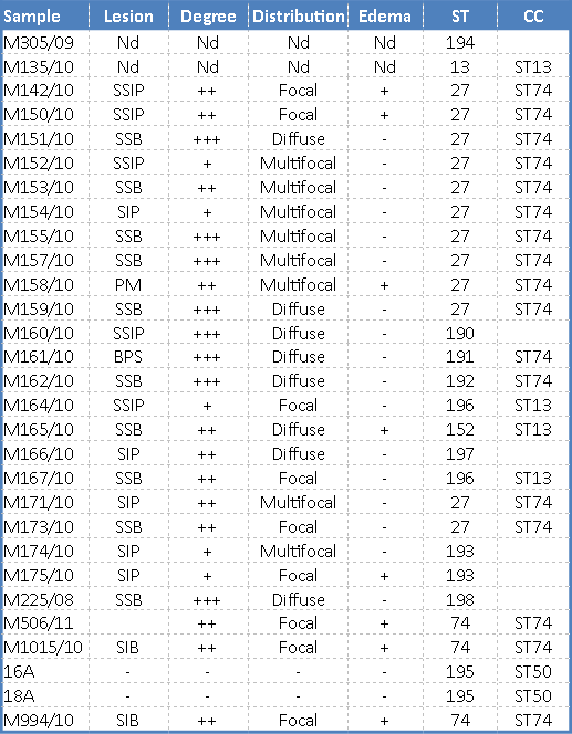 | Table 3- Microscopic lesions in histological analisys and MLST ST of P. multocida isolated from pig lung. |
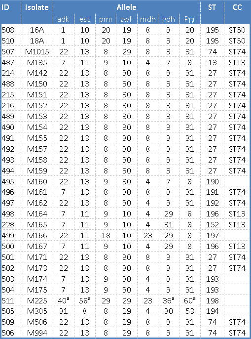 | Table 4-Genotype classification based on MLST. |