


ISSN : 0976-996X
EISSN : 0976-9978



ROPERTO SANTE1*, BORZACCHIELLO GIUSEPPE2, PERILLO ANTONELLA3, BALINT EMILIA4, LEONARDI LEONARDO5, DELLA SALDA LEONARDO6, FERRARI ANGELO7, IOVANE VALENTINA8, URRARO CHIARA9, LUCÀ ROBERTA10, RUSSO VALERIA11, MARCUS IOAN12, SALVATORE GERARDO13, ROPERTO FRANCO14
1Department of Pathology and Animal Health, Divisions of Infectious Diseases and General Pathology, Faculty of Veterinary Medicine, via Delpino 1, Naples University Federico II, 80137 Naples, Italy
2Department of Pathology and Animal Health, Divisions of Infectious Diseases and General Pathology, Faculty of Veterinary Medicine, via Delpino 1, Naples University Federico II, 80137 Naples, Italy
3Department of Health and Animal Welfare, Faculty of Veterinary Medicine, SP Casamassima Km 3, University of Bari, 70010 Valenzano, Italy
4Department of Internal Medicine, Faculty of Veterinary Medicine, 105 Independentei Street, University of Bucharest, Bucharest, Romania
5Department of Biopathological Sciences, Faculty of Veterinary Medicine, via S. Costanzo 4, University of Perugia, 06126 Perugia, Italy
6Department of Comparative Biomedical Sciences, Viale Crucioli, 122 University of Teramo, 64100 Teramo, Italy
7Center of Excellence on Comparative Oncology Research, Piazza Borgopila 39 - 10166 Genova, Italy
8Department of Pathology and Animal Health, Divisions of Infectious Diseases and General Pathology, Faculty of Veterinary Medicine, via Delpino 1, Naples University Federico II, 80137 Naples, Italy
9Department of Pathology and Animal Health, Divisions of Infectious Diseases and General Pathology, Faculty of Veterinary Medicine, via Delpino 1, Naples University Federico II, 80137 Naples, Italy
10Department of Pathology and Animal Health, Divisions of Infectious Diseases and General Pathology, Faculty of Veterinary Medicine, via Delpino 1, Naples University Federico II, 80137 Naples, Italy
11Department of Pathology and Animal Health, Divisions of Infectious Diseases and General Pathology, Faculty of Veterinary Medicine, via Delpino 1, Naples University Federico II, 80137 Naples, Italy
12Department of Pathophysiology, Faculty of Veterinary Medicine, Manastur Street, 3-5, Cluj, Romania
13Department of Health, Veterinary and Food Hygiene Division, Regione Basilicata, via Verrastro 9, 85100 Potenza, Italy
14Department of Pathology and Animal Health, Divisions of Infectious Diseases and General Pathology, Faculty of Veterinary Medicine, via Delpino 1, Naples University Federico II, 80137 Naples, Italy
* Corresponding Author : sante.roperto@unina.it
Received : 08-10-2011 Accepted : 17-11-2011 Published : 15-12-2011
Volume : 2 Issue : 2 Pages : 30 - 38
Vet Sci Res 2.2 (2011):30-38
DOI : http://dx.doi.org/10.9735/0976-996X.2.2.30-38
Conflict of Interest : None declared
Twenty-eight urothelial tumours of the urinary bladder were studied in 4-16-year-old cattle grazing on lands rich in bracken fern. Four primary adenocarcinomas, twelve papillary carcinomas, ten invasive carcinomas, two urothelial carcinomas with endophytic growth were examined. They were classified using morphological parameters which have been recently suggested in the report on the new histological classification of urothelial tumours of the urinary bladder of cattle. Furthermore, some incidental benign reactive processes such as cystitis glandularis and intestinal metaplasia, coexisting with urothelial tumours, were also investigated. E5 oncoprotein of bovine papillomavirus type 2 (BPV-2) was detected by immunoprecipitation in twenty-two tumours (ï¾79%). The remaining six tumours (ï¾21%) were E5-negative. Neuroendocrine differentiation was evident in fifteen tumours, four adenocarcinomas and eleven urothelial tumours; twelve of them, that is 80% (four adenocarcinomas and eight urothelial cancers) were E5-positive. Primary adenocarcinomas were composed of a number of multiple glands embedded in a loose stroma. Glandular lumina were lined by tall columnar cells which showed a marked immunoreactivity to chromogranin A, synaptophysin and serotonin. In eight papillary carcinomas containing varying degrees of a glandular or glandular-like differentiation, immunoreactivity was focally scattered among glandular structures. Two of them were E5-negative. Finally, immunoreactivity was also present in three invasive urothelial carcinomas. They were composed of glandular-like structures, cords and nests of neoplastic cells. Glandular-like structures showed a more marked immunoreactivity. A weak immunoreactivity was also seen in the cells at the edges of the nests. One of them appeared to be E5-negative. This study widens the spectrum of histological neoplastic urothelial lesions of cattle and affords further evidence that microscopic patterns of bovine urinary bladder tumours share striking morphological similarities with the human counterparts.
bovine papillomavirus type 2 (BPV-2), cattle, chronic enzootic haematuria, cystitis glandularis, intestinal metaplasia, neuroendocrine differentiation, urothelial carcinoma
Urinary bladder tumours are very rare in cattle, representing approximately 0.01% of all bovine malignancies [1] . Conversely, urinary bladder tumours are commonly encountered in adult cattle aged 4 or more years reared in hilly/mountain pasturelands rich in bracken fern (Pteridium spp.) [2-4] . The fern contains toxic principles and its prolonged ingestion, together with bovine papillomavirus type 2 (BPV-2) infection, plays a central role in bladder carcinogenesis [5-8] . However, the actual synergism between toxic principles of bracken fern and BPV-2 is still unclear. It has been suggested that BPV-2 is responsible for a latent infection of the urothelium that may result in bladder carcinogenesis since it can be activated by chemical carcinogens of bracken fern such as ptaquiloside (PT), known to be the major carcinogen of fern [4,9] . Urothelial carcinomas have a propensity for divergent differentiation leading to several variants, the histologic recognition of which is of crucial prognostic value since some of them may be associated with a different clinical outcome and may have a different therapeutic approach [10,11] . Neuroendocrine (NE) differentiation is a process observed in several tumours [12] . The spectrum of neoplastic lesions of the urinary bladder of cattle is becoming wider and wider [4,13] . Previous studies have shown that the histotypes of bovine urothelial tumours share striking morphological and biochemical features with their human counterparts which makes cattle a good model of bladder carcinogenesis [4,8,14-18] . The present paper focuses on some aspects of the focal NE differentiation observed in urothelial tumours and in some incidental tumour-like lesions of the urinary bladder in cattle grazing on bracken fern-infested lands. To our knowledge, NE differentiation has never been reported in urothelial tumours of the urinary bladder in veterinary comparative oncology.
Ethics Statement
In our cases we didn't perform any experimentation as we collected tissue samples in public slaughterhouses. All the animals we studied were slaughtered after a mandatory clinical ante-mortem examination, as required by the European Union legislation.
Samples of bladder neoplastic urothelium were collected at public slaughterhouses from twenty-eight 4- to 16-year-old cows that had been suffering from chronic enzootic haematuria for several years. All animals had been raised in hilly/mountain cattle households in the South of Italy and were known to have grazed on pastures rich in bracken fern. Normal bladder mucosa was obtained from five 2- to 10-year-old healthy cows which had grazed on pastures in which no bracken was present. Bladder samples were routinely divided into several parts.
One part was fixed in 10% neutral buffered formalin and processed routinely for paraffin embedding. Histologic diagnosis was assessed on 5µm sections stained with haematoxylin-eosin (HE) using morphological criteria suggested on the new, recently reported histological classification of urothelial tumours of the urinary bladder of cattle [4] . Another part was immediately frozen in liquid nitrogen, stored at -80° C until processed for molecular procedures. Furthermore, very small pieces (1 mm) were obtained from the remaining neoplastic part and were immediately fixed in 1% tannic acid-containing 2% glutaraldehyde in phosphate buffer. They were block-stained in 1% uranyl acetate in distilled water, dehydrated in graded alcohol and embedded in low viscosity Spurr resin. Ultrathin sections from selected areas were stained with uranyl acetate and lead citrate and examined with a JEM 1011 Jeol transmission electron microscope.
For immunohistochemical studies, 4-μm-thick sections were deparaffinized and blocked for endogenous peroxidase in 0.3% H2O2 in methanol for 20 minutes. Antigen enhancement was performed by pretreating with microwave heating (twice for 5 minutes each at 525 W) in citrate buffer, pH 6.00. The tissue sections were incubated with a blocking solution (Protein Block Serum Free, Dako, Denmark) for 20 minutes at room temperature in a humidified chamber to inhibit non-specific staining. Immunostaining was performed using, overnight at +4°C in a humidified chamber, the following antibodies: a monoclonal mouse anti-chromogranin A, (Clone DAK-A3 Dako, Denmark) diluted at 1 in 100/200, a monoclonal mouse anti-serotonin, (Clone 5HT-H209, Dako, Denmark), diluted at 1 in 50/100, a monoclonal mouse anti-synaptophysin, (Clone SY38, Dako, Denmark), diluted at 1 in 10/20. Slides were washed three times with phosphate buffer solution (PBS). A commercial kit (Dako, LSABK0690, Dako Cytomation, Burlingame, CA, USA) containing peroxidase-conjugated streptavidin and a mixture of biotinylated anti-rabbit/anti-mouse immunoglobulins was used and sections were incubated for 20 minutes in a humidified chamber. Slides were washed three times with PBS and color development was obtained with 5-20 minutes of diaminobenzidine treatment. Sections were counterstained with haematoxylin. Primary antibodies were omitted and replaced by PBS in corresponding negative control sections.
The frozen samples of tumours and normal urinary bladder mucosa were homogenized in RIPA lysis buffer containing 50 mM Hepes pH 7.5, 150 mM sodium chloride (NaCl), 0.25% sodium deoxycholate, 1% Triton, 1 mM ethylenediaminetetraacetic acid (EDTA) as elsewhere reported [16] .
Two-colour immunofluorescence staining was performed to investigate the colocalization of E5 and some neurosecretory granule types. Briefly, the sections were rinsed in PBS, pre-incubated with normal donkey serum (dilution 1 in 20 for 30 min). The sections were overlaid with polyclonal sheep anti-E5 (a kind gift of Prof. MS Campo, Glasgow University) diluted at 1 in 50 overnight at 4°C in a humid chamber. A secondary antibody Alexa Fluor 488 donkey anti-sheep (Invitrogen, Molecular Probes) was applied for 1 h at room temperature. The sections were overlaid with a mouse monoclonal anti-chromogranin A antibody (Clone DakA3, Dako, Denmark) diluted at 1 in 50/100 overnight at 4°C. A secondary antibody Alexa Fluor 546 donkey anti-mouse (Invitrogen, Molecular Probes) was applied (dilution 1 in 100) for 1 h at room temperature.
After washing 3 times with PBS, the slides were mounted under aqueous medium (Sigma, Milan, Italy). A confocal laser scanning microscope LSM-510 (Zeiss, Göttingen, Germany) was used. Alexa Fluor 488 (Invitrogen, Vienna, Austria) was excited at 495 nm and detected via a 519 nm band pass filter. Alexa Fluor 546 (Invitrogen, Vienna, Austria) was excited at 556 nm and detected with a 573 nm band pass filter. Two-channel frame-by-frame multitracking was used for detection to avoid “cross-talk†signals. The individual frames were scanned separately, with appropriate installation of the optical path for excitation and emission of each scan according to the manufacturer’s instructions.
Four tumours were entirely composed of multiple glands lined by columnar cells and embedded in a loose stroma; therefore, they were diagnosed as pure well-differentiated adenocarcinomas [Fig-1] . In all of them, NE cells were diffusely seen to be scattered among the columnar glandular cells lining some lumina. They were also manifest in clusters of cells arranged around small very small glandular lumina forming lumina. Although the NE cells were immunoreactive for synaptophysin and serotonin, a stronger immunoreactivity was evident for Chromogranin A [Fig-2] .
Eight endoluminal tumours showed microscopic patterns characterized by a papillary appearance with minimal to severe variability in architectural and cytologic features. Varying degrees of true glandular spaces within the tumours were seen; therefore, they were diagnosed as papillary urothelial carcinomas with glandular differentiation [Fig-3A] . NE cells were seen scattered intermingled with glandular formations. All NE cells were characterized by a clear immunoreactivity for chromogranin A, synaptophysin, serotonin [Fig-3B] , [Fig-3C] , [Fig-3D] . Three invasive tumours were characterized by nests and cords of cells with marked variation in size and shape, inducing a pseudosarcomatous stromal reaction. Many nests were cystically dilated and acquired luminal spaces [Fig-4] . Incidental findings such as intestinal metaplasia and cystitis cystica co-existing with urothelial tumours were found. Both intestinal metaplasia composed of mucin-secreting goblet cells and florid nonintestinal type cystitis cystica with cystic dilation were characterized by the presence of NE cells immunoreactive for serotonin only [Fig-5A] , [Fig-5B] . BPV-2 E5 oncoprotein was detected by immunoprecipitation in four adenocarcinomas and in eight urothelial tumours [Fig-6] . Morphologically, E5 protein and Chromogranin A granules were found to be mostly co-localized in luminal cells with confocal scanning laser microscope [Fig-7] .
Ultrastructurally, numerous intracytoplasmic polymorphic electron-dense structures bound by a single membrane identified as neurosecretory granules and bundles of tonofilaments were showed in several cancer cells, the cell junctions of which were characterized by the presence of both typical and atypical desmosomes [Fig-8] . The latter were composed of poorly differentiated desmosomes being characterized by tonofilaments from the cytoplasm converging upon plaques but by the absence of both dense material and intermediate lines in intercellular gaps [Fig-8] , insert).
Urothelial carcinoma of the bladder is unique among epithelial carcinomas in its divergent pathways of tumorigenesis [19] . It is well known that urothelial cancers show a marked propensity for divergent differentiation resulting in a wide spectrum of phenotypic variants which quite often show an aggressive clinical course portending a poor prognosis [11] . Urothelial cells can undergo a differentiation process to become NE cells expressing several NE markers. NE differentiation in bladder tumours has four major manifestations: carcinoid tumours, considered to be well-differentiated NE carcinomas; small cell NE carcinomas (SCNEC) and large cell NE carcinomas (LCNEC), both types known to be poorly differentiated NE carcinomas, and focal NE differentiation [20,21] . NE differentiation of tumours has very rarely been reported in domestic animals [22] . It is exceedingly rare in epithelial cancers of cattle being the known reports limited to a thymic carcinoma in a calf [23] . Our study shows variable degrees of NE differentiation in the form of isolated, single or small clusters of NE cells scattered and intercalated among the glandular structures in ï¾ 54% of the examined bladder tumours. In all adenocarcinomas, scattered NE cells lining lumina were diffusely seen. A marked NE differentiation was also seen in all the papillary and invasive urothelial carcinomas we examined, which were characterized by microscopic patterns containing glandular and glandular-like structures. How the spectrum of NE differentiation takes place in bladder carcinogenesis is unknown. It has been suggested that abnormal proliferation of NE cells originates from multipotential, undifferentiated or stem cell located in the urothelium [24,25] . The differentiation processes of the stem cells may be aberrantly regulated under pathological conditions [26] . Another hypothesis is that NE growth arises from neuroendocrine cells present within the normal urothelium or from an undefined population of submucosal neuroendocrine cells [27,28] . In most cases, NE cancer cells are admixed with conventional carcinoma including urothelial carcinoma, squamous cell carcinoma, adenocarcinoma, sarcomatoid carcinoma [10,29] . The coexistence of these epithelial components suggests that NE phenotype reflects divergent differentiation of multipotential urothelial cells [20] . Recently, genetic studies have shown that urothelial and small cell carcinoma components originate from the same cell in the urothelium [30,31] . Our ultrastructural investigations detected neurosecretory granules scattered in the cytoplasm of cancer cells containing prominent tonofilaments and showing classic desmosomes as well as so-called “attenuated†or “poorly differentiated†desmosomes considered to be of diagnostic value in tumours. These ultrastructural features allow us to suggest that NE cells are of epithelial origin and therefore are from multiphenotypic differentiation of urothelial cells. It is worthwhile remembering that the presence of desmosomes virtually guarantees that one is looking at epithelium, mesothelium and meninges or their tumours [32] .
In man, it has been shown that NE differentiation in bladder cancers is restricted to glandular and pseudoglandular lesions including glandular cystitis, intestinal metaplasia and primary adenocarcinoma [25,33] . Our findings are consistent with results reported in human medicine. Furthermore, we showed stronger NE differentiation in the glandular lumina both in urothelial and non-urothelial tumours.
Urothelial cancer cells can undergo a multiphenotypic differentiation process, after carcinogenesis is initiated, to become glandular cells [34] , and NE cells which acquire a phenotype like normal NE cells and express several NE markers. Similar NE differentiation has been shown in prostate adenocarcinoma cells [26,35] . Multipathways are known to be involved in regulating this process. It has been shown that the activation of c-Src upregulates specific transcriptional factors responsible for acquiring the NE phenotype [36] . More recently, it has been shown that Wnt-11, a protein known to modulate cell growth and differentiation as well as mitochondrial manganese superoxide dismutase (MnSOD), are essential inducers of NE differentiation in some cancers [12,37,38] .
The biological significance of NE differentiation has not well understood [37] . Very recently, it has been shown that serotonin, known to have powerful mitogen activity, promotes tumour growth by activating downstream targets of mTOR [39] . Furthermore, it has been suggested that chromogranin A is a modulator of the tumour microenvironment and its abnormal secretion could play important roles in tumour progression [40] . It is conceivable to believe that similar pathways can take place also in urothelial tumours of cattle.
We did not perform any specific and direct investigation about the potential role of papillomavirus infection in NE differentiation. However, we do not exclude the possibility that some BPV-2 proteins can be involved in divergent differentiation of urothelial tumours. It is worth noting that we have shown the oncoprotein E5 in a large number (12 out of 15) of bladder tumours with NE differentiation. Furthermore, E5 protein and neurosecretory granules appear to be co-localized in numerous cancer cells. It has been shown that c-Src, known to play an important role in NE cell differentiation of cancer cells [36] , can be activated also by E5 oncoprotein [41] . Furthermore activation of Wnt signaling pathway appears to occur in papillomavirus infection [42,43] . Further studies are required to better understand the role, if any, of papillomavirus infection in NE differentiation of the urothelial tumours in comparative oncology mostly in the light of the more and more emerging aetiological role of human papillomavirus infection in bladder carcinogenesis [44,45] .
This work was supported in part by grants from the Ministero Italiano dell’Università e della Ricerca Scientifica (MIUR), the Ministero Italiano delle Politiche, Agricole, Agroalimentari e Forestali (MIPAAF), the Assessorato alla Sanità della Regione Basilicata and Regione Campania. The authors thank Dr G. Di Domenico, Dr R. La Rizza, Dr G. Marino, Dr G. Massari, Dr G. Pizzolante, Dr A. Russillo, Dr G. Toma and Mr. F. Luposella for helping them to collect the cases. The authors declared that they had no conflicts of interest with respect to their authorship or the publication of this article.
[1] Meuten D.J. (2002) Tumors in Domestic Animals, DJ Meuten, Ed, Iowa State Press, Ames, pp 524-546.
» CrossRef » Google Scholar » PubMed » DOAJ » CAS » Scopus
[2] Pamukcu A.M., Price J.M. and Bryan G.T. (1976) Veterinary Pathology, 13, 110-122.
» CrossRef » Google Scholar » PubMed » DOAJ » CAS » Scopus
[3] Özkul I.A. and Aydin Y. (1996) British Veterinary Journal, 152, 473-475.
» CrossRef » Google Scholar » PubMed » DOAJ » CAS » Scopus
[4] Roperto S., Borzacchiello G., Brun B., Leonardi L., Maiolino P. et al. (2010) Journal of Comparative Pathology, 142, 95-108.
» CrossRef » Google Scholar » PubMed » DOAJ » CAS » Scopus
[5] Campo M.S., Jarrett W.F.H., Barron R., O’Neill B.W. and Smith K.T. (1992) Cancer Research, 52, 6898-6904.
» CrossRef » Google Scholar » PubMed » DOAJ » CAS » Scopus
[6] Borzacchiello G., Iovane G., Marcante M.L., Poggiali F., Roperto F. et al. (2003) Journal of General Virology, 84, 2921-2926.
» CrossRef » Google Scholar » PubMed » DOAJ » CAS » Scopus
[7] Borzacchiello G. and Roperto F. (2008) Veterinary Research, 39, 45-63.
» CrossRef » Google Scholar » PubMed » DOAJ » CAS » Scopus
[8] Roperto S., Brun R., Paolini F., Urraro C., Russo V. et al. (2008) Journal of General Virology, 89, 3027-3033.
» CrossRef » Google Scholar » PubMed » DOAJ » CAS » Scopus
[9] Campo M.S. (1997) Bovine papillomavirus and cancer. Veterinary Journal 154, 175-188.
» CrossRef » Google Scholar » PubMed » DOAJ » CAS » Scopus
[10] Epstein J.L., Amin M.B. and Reuter V.R. (2004) Bladder biopsy interpretation. Lippincott Williams & Wilkins, Philadelphia, pp 126-128.
» CrossRef » Google Scholar » PubMed » DOAJ » CAS » Scopus
[11] Shanks J.H. and Iczkowski K.A. (2009) Histopathology, 54, 885-900.
» CrossRef » Google Scholar » PubMed » DOAJ » CAS » Scopus
[12] Quiros-Gonzales I., Sainz R.M., Hevia D. and Mayo J.C. (2011) Free Radical Biology&Medicine, 50, 525-536.
» CrossRef » Google Scholar » PubMed » DOAJ » CAS » Scopus
[13] Brun R., Urraro C., Medaglia C., Russo V., Borzacchiello G. et al. (2008) Journal of Comparative Pathology, 139, 121-125.
» CrossRef » Google Scholar » PubMed » DOAJ » CAS » Scopus
[14] Roperto S., Ambrosio V., Borzacchiello G., Galati P., Paciello O. et al. (2005) Veterinary Pathology, 42, 812-818.
» CrossRef » Google Scholar » PubMed » DOAJ » CAS » Scopus
[15] Roperto S., Borzacchiello G., Casellato R., Galati P., Russo V. et al. (2007) Journal of Comparative Pathology, 137, 87-93.
» CrossRef » Google Scholar » PubMed » DOAJ » CAS » Scopus
[16] Roperto S., Borzacchiello G., Brun B., Costanzo F., Faniello M.C. et al. (2010) Journal of Comparative Pathology, 142, 9-18.
» CrossRef » Google Scholar » PubMed » DOAJ » CAS » Scopus
[17] Roperto S., Colabufo N.A., Inglese C., Urraro C., Brun R. et al. (2010) Journal of Comparative Pathology, 142, 19-26.
» CrossRef » Google Scholar » PubMed » DOAJ » CAS » Scopus
[18] Roperto S., De Tullio R., Raso C., Stifanese R., Russo V. et al. (2010) PLoS One, 5, e10299.
» CrossRef » Google Scholar » PubMed » DOAJ » CAS » Scopus
[19] Wu X.R. (2005) Nature Review Cancer 5, 713-725.
» CrossRef » Google Scholar » PubMed » DOAJ » CAS » Scopus
[20] Mazzucchelli R., Morichetti D., Lopez-Beltran A., Cheng L., Scarpelli M. et al. (2009) BJU International, 103, 1464-1470.
» CrossRef » Google Scholar » PubMed » DOAJ » CAS » Scopus
[21] Murali R., Kneale K., Lalak N. and Delprado W. (2006) Archives of Pathology & Laboratory Medicine, 130, 1693-1706.
» CrossRef » Google Scholar » PubMed » DOAJ » CAS » Scopus
[22] Ninomiya F., Suzuki S., Tanaka H., Hayashi S., Ozaki K. et al. (2008) Veterinary Pathology, 45, 181-187.
» CrossRef » Google Scholar » PubMed » DOAJ » CAS » Scopus
[23] Anjiki T. and Kadota K. (1999) Journal of Veterinary Medical Science, 61, 853-855.
» CrossRef » Google Scholar » PubMed » DOAJ » CAS » Scopus
[24] Blomjous C.E., Wos W., Schipper N.W., De Voogt H.J., Baak J.P. et al (1989) Journal of Clinical Pathology, 42, 1032-1039.
» CrossRef » Google Scholar » PubMed » DOAJ » CAS » Scopus
[25] Bollito E.R., Pacchioni D., Lopez-Beltran A., Volante M., Terrone C. et al. (2005) Analytical & Quantitative Cytology & Histology, 27, 218-224.
» CrossRef » Google Scholar » PubMed » DOAJ » CAS » Scopus
[26] Yuan T.C., Veeramani S. and Lin M.F. (2007) Endocrine-Related Cancer, 14, 531-547.
» CrossRef » Google Scholar » PubMed » DOAJ » CAS » Scopus
[27] Cramer S.F., Aikawa M., Cebelin M. (1981) Cancer, 47, 724-730.
» CrossRef » Google Scholar » PubMed » DOAJ » CAS » Scopus
[28] Oesterling J.E., Brendler C.B., Burgers J.K., Marshall F.F. and Epstein J.I. (1990) Cancer, 65, 1928-1936.
» CrossRef » Google Scholar » PubMed » DOAJ » CAS » Scopus
[29] Algaba F., Sauter G. and Schoenberg M.P. (2004) World Health Organization Classification of Tumours. Pathology and Genetics of Tumours of the Urinary System and Male Genital Organs, JN Eble, G Sauter, JI Epstein, IA Sesterhenn, Eds. IARC Press, Lyon, pp 135-136.
» CrossRef » Google Scholar » PubMed » DOAJ » CAS » Scopus
[30] Cheng L., Jones T.D., McCarthy R.P., Eble J.N., Wang M. et al. (2005) American Journal of Pathology, 166, 1533-1539.
» CrossRef » Google Scholar » PubMed » DOAJ » CAS » Scopus
[31] Abbosh P.H., Wang M., Eble J.N., Lopez-Beltran A., Mac Lennan G.T. et al. (2008) Modern Pathology 21, 355-362.
» CrossRef » Google Scholar » PubMed » DOAJ » CAS » Scopus
[32] Ghadially F.N. (1997) Ultrastructural pathology of the cell and matrix, FN Ghadially, Ed, Butterworth-Heinemann, Boston, pp 1184-1193.
» CrossRef » Google Scholar » PubMed » DOAJ » CAS » Scopus
[33] Williamson S.R., Lopez-Beltran A., Montironi R. and Cheng L. (2011) Histopathology, 58, 811-834.
» CrossRef » Google Scholar » PubMed » DOAJ » CAS » Scopus
[34] Kunze E. and Francksen B. (2002) Urology Research, 30, 66-78.
» CrossRef » Google Scholar » PubMed » DOAJ » CAS » Scopus
[35] Komiya A., Suzuki H., Imamoto T., Kamiya N., Nihei N. et al. (2009) International Journal of Urology, 16, 37-44.
» CrossRef » Google Scholar » PubMed » DOAJ » CAS » Scopus
[36] Yuan T.C., Veeramani S., Lin F.F., Kondrikou D., Zelivianski S. et al. (2006) Endocrine-Related Cancer, 13, 151-167.
» CrossRef » Google Scholar » PubMed » DOAJ » CAS » Scopus
[37] Quirós I., Sáinz R.M., Hevia D., Garcia-Suárez O., Astudillo A. et al. (2009) International Journal of Cancer, 125, 1497-1504.
» CrossRef » Google Scholar » PubMed » DOAJ » CAS » Scopus
[38] Uysal-Onganer P., Kawano Y., Caro M., Walker M.M., Diez S. et al. (2010) Molecular Cancer, 9, 55-65.
» CrossRef » Google Scholar » PubMed » DOAJ » CAS » Scopus
[39] Soll C., Jang J.H., Riener M.O., Moritz W., Wild J.P. et al. (2010) Hepatology, 51, 1244-1254.
» CrossRef » Google Scholar » PubMed » DOAJ » CAS » Scopus
[40] Corti A. (2010) Cellular and Molecular Neurobiology, 30, 1163-1170.
» CrossRef » Google Scholar » PubMed » DOAJ » CAS » Scopus
[41] Suprynowicz F.A., Baege A., Sunita I. and Schlegel R. (2002) Oncogene, 21, 1695-1706.
» CrossRef » Google Scholar » PubMed » DOAJ » CAS » Scopus
[42] Rampias T., Boutati E., Pectasides E., Sasaki C., Kountourakis P. et al. (2010) Molecular Cancer Research, 8, 433-443.
» CrossRef » Google Scholar » PubMed » DOAJ » CAS » Scopus
[43] Smeets SJ, van der Plas M, Schaaij-Visser TB, van Veen EA, van Meerloo J et al. (2011) International Journal of Cancer, 128, 1596-1605.
» CrossRef » Google Scholar » PubMed » DOAJ » CAS » Scopus
[44] Shigehara K, Sasagawa T, Doorbar J, Kawaguchi S, Kobori Y et al. (2011) Journal of Medical Virology, 83, 277-285.
» CrossRef » Google Scholar » PubMed » DOAJ » CAS » Scopus
[45] Shigehara K., Sasagawa T., Kawaguchi S., Nakashima T., Shimamura M. et al. (2011) Cancer, 117, 2067-2076.
» CrossRef » Google Scholar » PubMed » DOAJ » CAS » Scopus
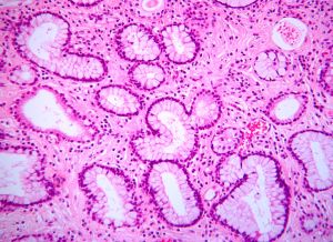 | Fig. 1- Adenocarcinoma of the urinary bladder. Numerous glandular structures lined by cuboidal and columnar cells are embedded in a loose stroma. HE x220. |
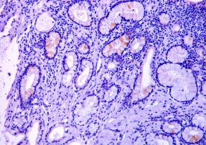 | Fig. 2- Adenocarcinoma of the urinary bladder. Numerous columnar cells manifest an evident positivity for chromogranin A. Streptavidin-biotin complex method. Haematoxylin counterstain x220. |
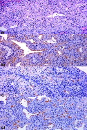 | Fig 3. (a) Several lumina are evident in a papillary urothelial carcinoma with glandular differentiation. HE. (b) Papillary urothelial carcinoma with glandular differentiation. Many cells of gland lumina are immunoreactive for chromogranin A. Streptavidin-biotin complex method. Haematoxylin counterstain. (c) Papillary urothelial carcinoma with glandular differentiation. Some cells lining gland lumina show an immunoreactivity for serotonin. Streptavidin-biotin complex method. Haematoxylin counterstain. (d) Papillary urothelial carcinoma with glandular differentiation. Many glandular structures are lined by cells immunoreactive for synaptophysin. Streptavidin-biotin complex method. Haematoxylin counterstain x220. |
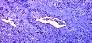 | Fig. 4- Invasive high-grade urothelial carcinoma. An invasive urothelial carcinoma composed of cystically dilated neoplastic nests with luminal space is manifest. A strong pseudosarcomatous reaction of the stroma is also evident. Some cells of luminal space are immunoreactive for chromogranin A. Streptavidin-biotin complex method. Haematoxylin counterstain x220 |
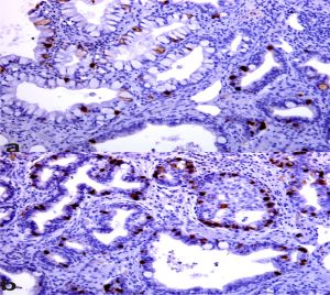 | Fig. 5- (a) Intestinal metaplasia composed of goblet cells immunoreactive for serotonin is shown in urinary bladder. Streptavidin-biotin complex method. Haematoxylin counterstain. (b) Florid cystitis cystica with cystic dilation composed of cuboidal cells immunoreactive for serotonin is evident. Streptavidin-biotin complex method. Haematoxylin counterstain x220 |
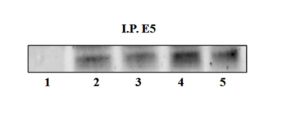 | Fig. 6- Oncoprotein E5 detected by immunoprecipitation in bladder lesions. Lane 1 – Healthy cow; lanes 2-5: neoplastic |
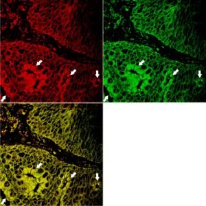 | Fig. 7- Two-colour immunofluorescence labeling demonstrates co-localization of E5 and Chromogranin A. Luminal cells show colocalization (yellow) of E5 (green; Alexa Fluor 488) and Chromogranin A (red, Alexa Fluor 546) (white arrows). x220 |
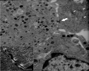 | Fig. 8- Ultrastructural neurosecretory granules, bundles of tonofilaments (thin arrows) and desmosomes (large arrow) are evident in and between cancer cells. x12,000. Many desmosomes appear to be poorly differentiated being composed of tonofilaments converging upon plaques but lacking in dense material and intermediate lines in the intercellular gaps (insert, x12,000). |