


ISSN : 2249-9466
EISSN : 2249-9474



SANGHAVI J.B.1*, BHOYAR K.K.2, GAWANDE U.H.3
1Department of Computer Science and Engineering, Yeshwantrao Chavan College of Engineering Nagpur, India.
2Department of Information Technology, Yeshwantrao Chavan College of Engineering Nagpur, India.
3Department of Information Technology, Yeshwantrao Chavan College of Engineering Nagpur, India.
* Corresponding Author : sjignyasa8588@gmail.com
Received : 21-02-2012 Accepted : 15-03-2012 Published : 19-03-2012
Volume : 2 Issue : 1 Pages : 22 - 24
Adv Med Informat 2.1 (2012):22-24
Content based image retrieval (CBIR) is the application of computer vision. It is the technique to the image retrieval problem also known as query by image content. CBIR is the major topic of research in medical domain due to increase in large medical image databases. Instead of text based searching, CBIR efficiently retrieves images that are visually similar to query image. CBIR allows the retrieval of similar images based on features directly extracted from the input image. This paper will focus on recent advances in CBIR systems in medical domain. It also focuses on the various feature extraction techniques and algorithms implemented for CBIR systems in different cases of medical domain.
expert system, CBIR (Content based image retrieval) systems, automated system, medical CBIR, image retrieval.
Medical images like MRI images, CT scan images, etc. have become a main investigation tool for medical diagnosis. Digital imaging is becoming the standard for all image acquisition devices. With the generalization of digital acquisition, there is an increasing need for data storage and retrieval. Medical images represent an enormous amount of data: the annual production of a single average size radiology department represents tens of terabytes of data. Therefore, petabytes of medical images are produced in industrialized countries each year. For such huge data there is a need of proper storing and retrieval system. CBIR system is found to be very efficient retrieving system.
Content Based Image Retrieval (CBIR) has emerged in the early 1990s. The principal aim is to represent each image as a feature vector and to measure the similarity between the image and image database and to retrieve similar digital images based on features and not on textual annotations. In CBIR query is given in the form of image. Then image database is searched through all images in order to find those with the most similar indices which are returned as the images most alike to the query image. [Fig-1] presents the general Architecture of CBIR systems proposed in [1] .
Many CBIR were developed during the last years, both by commercial firms and academia. The earliest and most famous one is QBIC (Query By Image Content) [2] which was proposed by IBM. This system only used low level features - mainly color, texture and shape - and did not include any semantic level. Later systems like Blobworld [3] include a segmentation step in the query process to integrate higher level information. In Blobworld, the segmentation step is based on color, position and texture features and leads to a small number of homogeneous regions called blobs. Submitting a request, the user can select the relevant blobs he wants the query to be applied on.
Despite various CBIR developments, medical images in different fields are very particular and require a specific design of CBIR systems. There exists a large number of medical image acquisition devices among which computed tomography scanners (CT), magnetic resonance imagers (MRI), ultrasound probes (US) and nuclear imagers are the most widely used. They provide images with very different properties in terms of resolution, contrast, and signal to noise ratio. They are highly specialized and they produce images giving very different information on the human body anatomy and physiology. A. Kak and C. Pavlopoulou notice that medical image retrieval must often be processed according to pathology bearing regions which are precisely delimited on the images and could not automatically detected in the general case. [4] Moreover, low level features like color, texture or shape are not sufficient to describe medical images [1] . As a consequence, medical CBIRs require a high level of content understanding and interpretation of images, which implies their automatic segmentation [5] . Finally, a high level of query completion and accuracy is required by such systems to make them reliable from a clinical point of view [6] .
B.F. Jones, G. Schaefer and S.Y. Zhu proposed a generic approach for the automated processing of medical infrared images. In recent years, there has been a resurgence of interest in the application of infrared thermal imaging in medicine due to improvements in camera technology and the promise of reduced costs. Thermography captures the natural thermal radiation generated by an object at a temperature above absolute zero. The radiance from human skin is an exponential function of the surface temperature, which in turn is an indicator of the level of blood perfusion in the skin. Changes in blood perfusion may occur for a variety of reasons, such as inflammation, angiogenesis, and previous traumas. It is well know that asymmetrical temperature distributions and hot and cold spots are strong indicators of an underlying dysfunction [7] .
Dr. B G Prasad and Krishna A N evaluated the performance of two statistical methods of texture features proposed by Haralick and Tamura for retrieving similar cases for 2-D CT scan brain images. Lam.M et.al computed tomography (CT) image of a lung nodule as a query and retrieves similar lung nodule images based on content-based image features [16] .
A retrieval system for magnetic resonance images (MRI) of the brain has been reported in [8] . The main image feature that is used for characterizing these images is the shape of the ventricular region. In another system reported in [10] , the images in the database consist of a single tumor in the center without any background texture. The system presented in [11] aims at aiding physicians in the diagnosis of lymph proliferative disorders of the blood.
Cai et al. describes in [12] a CBIR system for positron emission tomographic (PET) images of the brain. In this case, a set of physiological features as well as text are used for retrieval.
The ASSERT system reported by [13] is designed for high resolution computed tomography (HRCT) images of the lung where a rich set of textural features derived from the disease-bearing regions is important for the characterization of the images. The physician is an integral part of ASSERT, in the sense that it’s the physician who delineates the PBR's when an image is entered into the database and, also, in the query image. A. Kak and C. Pavlopoulou focus on the main contributions of ASSERT regarding HRCT lung images as well as some more recent work on HRCT liver images [4] .
Ke Chen et. al proposed a hierarchical retrieval method for ultrasound images. In this color feature and texture feature are considered [14] . C.I. Christodoulou et.al developed an automated image retrieval and classification system for the retrieval of similar carotid plaque ultrasound images, which will assist the physician in making his diagnostic decision based on similar previous cases. The neural self organizing map (SOM) and the statistical K-nearest neighbor (KNN) classifiers were used for the retrieval and the classification of the carotid plaques into symptomatic or asymptomatic. Twenty different feature sets including texture, shape, morphological, histogram and correlogram features were extracted from the carotid plaque images and the classification results were further combined in order to improve the success rate [17] .
Jaime-Castillo et.al, describes a Content-based image retrieval (CBIR) system to help in the diagnosis and treatment process of the scoliosis pathology. To do this, the system includes a module for the automatic extraction of scoliosis measures from X-rays [15] .
Lei Zheng et.al proposed a content-based image retrieval system employing client/server architecture to access supercomputing power from the physician’s desktop. The system retrieves images and their associated annotations from a networked microscopic pathology image database based on content similarity to user supplied query images. They evaluated the similarity based on four image feature types: color histogram, image texture, Fourier coefficients and wavelet coefficients, using the vector dot product as a distance metric [19] . James Z. Wang et.al developed Pathfinder, an efficient multiresolution region-based searching system for high-resolution pathology image libraries. The system uses wavelets and the IRM (Integrated Region Matching) distance [20] . D. Comaniciu et.al proposed a system to assist the physicians in differential diagnosis of lymphoproliferative disorders of blood cells from digitized specimens. They described a task-oriented indexing and retrieval system which exploits the informational content of digitized specimens in order to assist pathologists to discriminate among Chronic Lymphocytic Leukemia (CLL), Follicular Center Cell Lymphoma (FCC), Mantle Cell Lymphoma (MCL) and healthy leukocytes (NORMAL) [11] . J.Sanghavi et.al proposed content based image retrieval system for microscopic blood peripheral images of blood related diseases. The [Fig-2] shows the architecture of CBIR for pathology images [21] .
The huge amount of visual data produced in medical domain increased the importance of developing new and alternative access methods to complement it. Content based Image Retrieval has become an important area of research in medical domain also. The large number of research publications in the field of CBIR systems especially in recent years shows that it is very active research field and is getting more attention. This will hopefully advance the field, as new tools and technologies will be developed and performance will increase. In this paper we have highlighted the maximum areas in which CBIR is developed.
[1] Lehmann T., Wein B., Dahmen J., Bredno J., Vogelsang F. and Kohnen M. International Society for Optical Engineering (SPIE), 3972(32).
» CrossRef » Google Scholar » PubMed » DOAJ » CAS » Scopus
[2] Flickner M., Sawhney H., Niblack W., Ashley J., Huang Q., Dom B., Gorkani M., Hafner J., Lee D., Petkovic D., Steele D. and Yanker P. IEEE Computer.
» CrossRef » Google Scholar » PubMed » DOAJ » CAS » Scopus
[3] Carson C., Thomas M., Belongie S., Hellerstein J. and Malik J. (1999) Third International Conference on Visual Information Systems, 1614, 509-516.
» CrossRef » Google Scholar » PubMed » DOAJ » CAS » Scopus
[4] Kak A. and Pavlopoulou C. (2002) Visualization, Transmission.
» CrossRef » Google Scholar » PubMed » DOAJ » CAS » Scopus
[5] Muller H., Michoux N., Bandon D. and Geissbuhler A. (2004) International Journal of Medical Informatics, 73(1), 1.
» CrossRef » Google Scholar » PubMed » DOAJ » CAS » Scopus
[6] Tagare H.D., Jane C.C. and Duncan J. (1997) J Am Med Inform Assoc, 4(3), 184, 198.
» CrossRef » Google Scholar » PubMed » DOAJ » CAS » Scopus
[7] Jones B.F., Schaefer G. and Zhu S.Y. (2004) 26th Annual International Conference of the IEEE EMBS.
» CrossRef » Google Scholar » PubMed » DOAJ » CAS » Scopus
[8] Chu W.W., Hsu C.C., Cardenas A.F. and Taira R.K. (1998) IEEE Trans. on Knowledge and Data Engineering, 10(6), 872-888.
» CrossRef » Google Scholar » PubMed » DOAJ » CAS » Scopus
[9] Hsu C.C., Chu W.W. and Taira R.K. (1996) IEEE Trans. On Knowledge and Data Engineering, 8(4), 522-532.
» CrossRef » Google Scholar » PubMed » DOAJ » CAS » Scopus
[10] Korn F., Sidiropoulos N., Faloutsos C., Siegel E. and Protopapas Z. (1998) IEEE Trans. on Knowledge and Data Engineering, 10(6), 889-904.
» CrossRef » Google Scholar » PubMed » DOAJ » CAS » Scopus
[11] Comaniciu D., Meer P., Foran D. and Medl A. (1998) Workshop on Applications of Computer Vision. IEEE.
» CrossRef » Google Scholar » PubMed » DOAJ » CAS » Scopus
[12] Cai W., Feng D. and Fulton R. (2000) IEEE Transactions on Information Technology in Biomedicine, 4(2), 152-158, 2000.
» CrossRef » Google Scholar » PubMed » DOAJ » CAS » Scopus
[13] Shyu C., Brodley C., Kak A., Kosaka A., Aisen A. and Broderick L. (1999) Computer Vision and Image Understanding, 75(1/2), 111-132.
» CrossRef » Google Scholar » PubMed » DOAJ » CAS » Scopus
[14] Ke Chen, Jiangli Lin, Yuanwen Zou, Guangfu Yin (2009) Content-based Medical Ultrasound Image Retrieval Using a Hierarchical Method, IEEE.
» CrossRef » Google Scholar » PubMed » DOAJ » CAS » Scopus
[15] Jaime-Castillo S., Medina J.M., and Sanchez D. IEEE International Conference on Fuzzy systems.
» CrossRef » Google Scholar » PubMed » DOAJ » CAS » Scopus
[16] Lam M., Disney T., Pham M., Raicu D. and Furst J. (2007) SPIE Medical Imaging Conference.
» CrossRef » Google Scholar » PubMed » DOAJ » CAS » Scopus
[17] Christodoulou C.I., Pattichis C.S., Kyriacou E., Nicolaides A. (2010) Image Retrieval and Classification of Carotid Plaque Ultrasound Images.
» CrossRef » Google Scholar » PubMed » DOAJ » CAS » Scopus
[18] Prasad B.G. and Krishna A.N. IEEE .
» CrossRef » Google Scholar » PubMed » DOAJ » CAS » Scopus
[19] Lei Zheng, Wetzel A.W., John Gilbertson and Becich M.J. IEEE Transaction on Information Technology in Biomedicine, 7(4).
» CrossRef » Google Scholar » PubMed » DOAJ » CAS » Scopus
[20] Wang J.Z., Math M.S. (2000) AMIA Symposium.
» CrossRef » Google Scholar » PubMed » DOAJ » CAS » Scopus
[21] Sanghavi J., et al. (2012) ITAC.
» CrossRef » Google Scholar » PubMed » DOAJ » CAS » Scopus
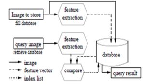 | Fig. 1- General Architecture of CBIR systems |
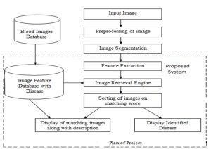 | Fig. 2- CBIR architecture for Pathology images |