


ISSN : 0976-996X
EISSN : 0976-9978



EZEQUIEL M.C.1, FARIA A.C.S.2, PAULA D.A.J.3, PITCHENIN L.C.4, BRANDÃO L.N.S.5, DE SOUZA R.L.6, NAKAZATO L.7, DUTRA V.8*
1Department of Veterinary Clinical Sciences, Av. Fernando Corrêa da Costa, nº 2367, Bairro Boa Esperança. Cuiabá, MT, 78060-900, Brazil.
2Department of Veterinary Clinical Sciences, Av. Fernando Corrêa da Costa, nº 2367, Bairro Boa Esperança. Cuiabá, MT, 78060-900, Brazil.
3Department of Veterinary Clinical Sciences, Av. Fernando Corrêa da Costa, nº 2367, Bairro Boa Esperança. Cuiabá, MT, 78060-900, Brazil.
4Department of Veterinary Clinical Sciences, Av. Fernando Corrêa da Costa, nº 2367, Bairro Boa Esperança. Cuiabá, MT, 78060-900, Brazil.
5Department of Veterinary Clinical Sciences, Av. Fernando Corrêa da Costa, nº 2367, Bairro Boa Esperança. Cuiabá, MT, 78060-900, Brazil.
6Department of Veterinary Clinical Sciences, Av. Fernando Corrêa da Costa, nº 2367, Bairro Boa Esperança. Cuiabá, MT, 78060-900, Brazil.
7Department of Veterinary Clinical Sciences, Av. Fernando Corrêa da Costa, nº 2367, Bairro Boa Esperança. Cuiabá, MT, 78060-900, Brazil.
8Department of Veterinary Clinical Sciences, Av. Fernando Corrêa da Costa, nº 2367, Bairro Boa Esperança. Cuiabá, MT, 78060-900, Brazil.
* Corresponding Author : valdutra@ufmt.br
Received : 30-01-2012 Accepted : 15-03-2012 Published : 18-10-2012
Volume : 3 Issue : 1 Pages : 48 - 54
Vet Sci Res 3.1 (2012):48-54
DOI : http://dx.doi.org/10.9735/0976-996X.3.1.48-54
Conflict of Interest : None declared
Osteochondrosis (OC) is seen as the main cause of leg weakness in pigs. Its etiology and pathogenesis is not fully understood, but any abnormalities in the formation of hypertrophic chondrocytes and disrupted blood supply to the growth cartilage are predisposing factors. The aim of this study was to characterize the changes in transcript profiles of joints from OC and non OC-affected pigs by the Representational Difference Analysis (RDA) technique. From both RDA libraries 11 sequencing reads had quality analysis. Quantitative real-time analysis was performed to examine the expression levels of HBB, COL1A1 and TPP1 genes. The expression of HBB and TPP1 genes were up regulated in the OC joint. There was no difference in expression of COL1A1 gene between healthy and OC joints. In this study the finding genes might play some role in development of OC in pigs. However, our findings should lead to further investigation of their implications.
Gene expression, Osteochondrosis, Representational Difference Analysis.
Osteochondrosis (OC) is a common and clinically important disease of growth cartilage that occurs in domestic animals as well as in humans (Bohndorf, 1998 Yamagiwa, 2010). In pork production, this disease often referred to as ‘leg weakness’, in addition to causing economic losses, affect animal welfare (Jorgensen, 2000; Kadarmideen et al. 2004; Guo et al. 2009).
The disease occurs at high frequencies in growing pigs in the most commercial breeds (Uhlhorn et al., 1995), and the lesions are commonly found in the medial aspect and the sagital ridge of the distal condyle of the humerus and the medial condyle of the femur (Nakano et al. 1987; Alberton, 2007).
Chondrocytes in growth cartilage during the process of endochondral ossification, undergo proliferation followed by several steps of maturation, culminating in hypertrophy and physiological death (Mackie et al. 2008). The OC is characterized by a disturbance in the normal cellular differentiation of cartilage cells in growth, failure occurring in the process of endochondral ossification (Ekman and Carlson, 1998; Semevolos et al. 2005). The series of events that occurs during this process is regulated by a number of secreted proteins, signalling cascades and transcription factors (Mackie et al. 2008). Although many factors including genetic, trauma, hormonal disorders, ischemia and anatomic conformation appear to play an important role in the development of the disease (Kadarmideen et al. 2004; Ytrehus et al. 2007), its etiology and pathogenesis is not well understood yet (Grondalen, 1974; Laenoi et al. 2010).
Studies on signal transduction and gene expression in swine cartilage metabolism and disease are limited. Studies involving functional genomics approaches illustrate an increasing effort to identify the genetic basis of OC pathogenesis in different species (Wittwer et al. 2008; Laenoi et al. 2011). Gene expression analysis from OC-affected horses have been made and identified changes in gene expression of a number of genes. In addition, metabolic pathways analysis showed dysregulation of several signaling pathways related to cartilage formation or repair (Mirams et al. 2009; Serteyn et al. 2010).
The aim of this study was to characterize the changes in transcript profiles of joints from OC and non OC-affected pigs through the Representational Difference Analysis (RDA) technique.
Healthy and OC joints from the articular surface to the bone from proximal femur bone from swine were excised at the slaughterhouses under federal inspection. For the external evaluation of articular cartilage and the cut surfaces of the extremities, the Kirk et al. (2008) modified criteria was used: (1) erosion: Articular cartilage reduction, (2) ulceration: articular cartilage absence with subchondral bone exposure and flaps, (3) repair: connective tissue or fibrocartilage is formed, (4) osteophytes: bony projections on the articular cartilage, (5) retraction: protusion of the articular cartilage over subchondral bone, (6) normal: no changes in the articular cartilage. The samples were divided into two parts, for RNA extraction and histopathology. Samples for RNA extraction were kept snap-frozen in liquid nitrogen until the processing. Samples for histolopathology were fixed in a solution of 10% buffered formalin for 24 to 48 hours.
The samples were subjected to a demineralization process with nitric acid to 8% for up to 96 hours. Histology sections were prepared in order to observe all the structural elements of a bone extremity: articular cartilage, growth plate and epiphyseal and metaphyseal trabecular bone.
For microscopic evaluation were performed hematoxylin-eosin (HE) and Masson’s Trichrome staining in thin sections of the samples (Kammerman et al. 1992).
Total RNA was isolated and cDNA was extracted from healthy and OC joint according to the Clements et al. (2006) protocol and SMART PCR synthesis kit (Clontech Laboratories), respectively, according to manufacturer’s instructions. RNA concentration was ascertained spectrophotometrically (Epoch, Biotek) at 260/280 nm and its integrity was checked by agarose gel electrophoresis. First-strand cDNA synthesis was performed with reverse transcriptase Superscript II (Invitrogen). An aliquot of 2 μl of first-strand cDNA was used as template for second-strand synthesis.
Representational Difference Analysis (RDA) was performed according to a modified protocol described by Dutra et al. (2004) for generation of differential expression libraries (Hubank and Schatz, 1994; Pastorian et al. 2000). A double-stranded cDNA sample (1 μg) of each condition (healthy and OC joint) was digested with Sau3AI (Invitrogen), the digestion products were purified using GFX (GE Healthcare) and analysed on 1% agarose gel.
To generate differential products two successive RDA rounds employing different adapters were done. In the first round 300 pmol of NBam24/12 adapters were ligated to the digested cDNA to be used as tester and hybridized to a 1:10 excess of driver cDNA (18 hours at 67 â°C). The first round of RDA generates the first differential product (PD1). To generate the second differential product (PD2), 300 pmol of JBam 24/12 adapters were ligated to the tester, and the tester/driver ratio was raised to 1:100. After each round, differentiaI products were digested with Sau3AI to remove the adapters that had been incorporated into cDNAs. In the RDA protocol, cDNA from OC joint served as the tester population and was hybridized to cDNA from healthy joint as driver, generating the library forward. When the healthy joint was used as tester and the OC joint as the driver, it was generated the reverse library. Oligonucleotides employed in RDA protocol are listed in [Table-1] .
An aliquot of PD2 was purified using GFX kit (GE Healthcare) and ligated into pGEM-T Easy Vector (Promega) according to the manufacturer’s instructions and Promega's pGEM T kit protocol. The inserts were transformed Escherichia coli XL1-Blue competent cells according to Sambrook and Russel (2001). After this step, the cloned inserts were extracted according to Sambrook and Russel (2001), purified using GFX (GE Healthcare) and sequenced with the Dyenamic ET Dye Terminator cycle sequencing kit for Megabace DNA analysis system using the MegaBace 1000 sequencer (GE Healthcare). The corresponding primers employed were the pGEM-T Easy vector (forward/reverse).
Read quality was checked and the reads were assembled by the Phred program (Ewing et al. 1998). In order to remove any segments of vector origin before sequence analysis, the differentially expressed sequence tags (ESTs) were screened using UniVec database at the National Center Biotechnology Information (NCBI). The ESTs resulting were compared to GenBank Database using the BLASTx program (Altschul et al. 1997) at the NCBI and PEDE (Pig Est Data Explorer). Sequences returning matches with an E-value ≤ 10â»Â¹â° and approximately 100% of sequence similarities were annotated and classified based on their putative molecular function and/or biological process using UniGene, Interpro and Pfam databases.
For analyses of a subset differentially expressed genes by quantitative real-time (RT-qPCR) analysis, gene-specific primers were designed using Oligo Explorer (Freeware, Teemu Kuulasmaa, Finland), the predict product length varied between 100 bp and 200 bp and are listed in [Table-1] . Real-time PCR assays were performed using StepOne Plus (Applied Biosystems). The PCR thermal cycling conditions were as follows: an initial denaturation at 95°C for 10 minutes and 40 cycles at 95°C for 15 seconds, 60°C for 60 seconds. SYBR green (Invitrogen) was used in the reaction, adding 5 pmol of each primer and 1μl of template cDNA at a final volume of 25 μl. All experiments were done in triplicate for each gene. Melting curve analysis was performed at the end of the reaction to confirm a single PCR product. The reference gene used as normal control was TATA-binding protein (TBP) (Laenoi et al. 2010), a gene encoding protein involved in transcriptional regulation (Pugh, 2000; Hochheimer and Tjian, 2003). Relative expression data was obtained using the 2ˉ∆∆CT method (Livak e Schmittgen, 2001).
Osteochondritis dissecans lesions were characterized by epiphyseal articular complex dysplasia causing cracks and retaining areas of cartilage due to necrosis areas having or not epiphyseal trabeculae microfractures. The joint collected showed retention areas of cartilage in the metaphyseal region, characterized by failure mineralization of hypertrophic chondrocyte columns [Fig-1] . It was also observed irregularities in the columns of chondrocytes in both proliferative and the hypertrophic zone, chondrolysis and condroclasia; eosinophilic streaks and areas of hypertrophic chondrocytes retention, epiphyseal plate-shaped cone even fiseais fractures [Fig-2] , osteonecrosis and trabecular microfractures.
The PD2 obtained after the second round of RDA showed a molecular weight profile below 1000 pb [Fig-3] . The cloning of this products into pGEM T Easy vector, resulted in 100 clones of approximately 400 bp. All clones were sent for sequencing and 11 had quality for analysis. The results of computational homology search for cDNAs obtained are shown in [Table-2] and [Table-3] . Of these 11 sequencing reads only 5 (3 from healthy joint library and 2 from OC joint library) passed the sequence quality analysis (Phred quality ≥ 30) and shows significant matches to genes in the GenBank database. They are listed in [Table-4] . The subtracted library generated from healthy joint contains genes encoding proteins responsible for oxygen transport such as α- and β-globin, both hemoglobin subunits encoded by HBA and HBB genes, respectively (Schechter, 2008). Another gene identified in this condition was the Collagen type I alpha 1 (COL1A1), which encodes the primary subunit of type I collagen, which forms more than 90% of the organic mass of bone and is a heterotrimer composed of two identical chains α1 (I) - and a β2 (I) - (Gelse et al. 2003). The subtracted library generated from OC joint contains a gene encoding a lysosomal proteinase (Tripeptidyl peptidase I - TPP1) related to protein degradation (Vines and Warbuton, 1998; Koike et al. 2002), and the gene HBB, also found in healthy joint subtracted library.
For further confirmatory data, quantitative real-time RT-qPCR analysis was performed to examine the expression of 3 genes selected for analysis, identifying using the RDA technique. The expression levels of HBB, COL1A1 and TPP1 were compared between healthy and OC joints. The HBB and TPP1 genes were up regulated in the OC joint compared with healthy joint. There was no difference in mRNA expression of COL1A1 between healthy and OC joints [Fig-4] .
Osteochondrosis is one of most serious problems in pig production (Laenoi et al. 2010). A clear understanding of OC pathogenesis is important to prevent and to improve disease resistance pigs, which is important to animal welfare in addition to improve production efficiency in animals and reducing losses due the disease.
Several studies have been done about the molecular mechanisms of genes possibly involved in OC pathogenesis (Mirams et al. 2009; Laenoi et al. 2010; Laenoi et al. 2011b). Genes which are involved in maintaining the normal structure of articular cartilage might be important factors in the pathogenesis of OC. A differential expression of these genes may lead a disturbance in the cartilage metabolism, which may trigger the disease (Laenoi et al. 2010). The gene products identified from the healthy and OC joint are involved in a variety of biological processes and encodes proteins that comprise structural components or catalytic function. The main focus of this study was on identifying genes with differential expression in healthy and OC joints from pigs. We investigated the gene expression between healthy and OC joints to explore a possible relationship between the candidate genes - previously identified by RDA technique - and OC occurrence in pigs.
The TTP1 gene, which was found to be up regulated at OC joint, encodes a lysosomal exopeptidase (Sleat et al. 1997). In humans, mutations in this gene are associated with an autosomal recessive lysosomal storage disorder called Classic late-infantile neuronal ceroid lipofuscinosis (LINCL) (Sleat et al. 1997; Rawlings and Barrett, 1999), which lead to massive accumulation of autofluorescent storage material in various tissues and organs. However, the central nervous system shows severe damage (Dekaban, 1978). Studies demonstrate that TPP1 is widely distributed in various tissues of mammals (Doebber et al. 1978; Watanabe et al. 1992; Vines and Warburton, 1998; Junaid et al. 2000; Du et al. 2001; Kida et al. 2001; Koike et al. 2002). Although it’s proteolytic activity did not always coincide with the expression levels of the protein (Koike et al., 2002).
Koike et al. (2002), studying the expression of TPP1 in various tissues of rats and mice by biochemical and immunohistochemichal techniques, showed that in osteoblasts and osteocytes in mouse tibia bone contained immunodeposits for TPP1, which became more intense in osteoclasts and chondrocytes of the articular cartilage (Koike et al. 2002).
TPP1 releases Gly-Pro-X triplets from synthetic collagen-like polymers, suggesting that this enzyme might be involved in earlier stages of collagen degradation (McDonald et al. 1985). Page et al. (1993) also suggest that TPP1, secreted by osteoclasts, is involved at the some stage in the degradation of bone collagen. Various studies have focused on the distribuction of proteinases that may be implicated in collagen metabolism in normal articular cartilage and in samples from osteochondrotic lesions (Hernandez-Vidal et al. 1997; Gläser et al. 2003). The functional role of TPP1 in the formation or maintenance of OC lesion at different stages of the disease, therefore, remains to be investigated.
The COL1A1 gene encodes two of the three subunits of type I collagen, the most abundant protein in mammals and the main structural protein of bones, teeth and tendons (Viguet-Carrin et al. 2006). No difference expression of COL1A1 between healthy and OC joint was observed by RT-qPCR. Although this gene have been detected just at healthy joint by RDA technique. Wardale and Duance (1994), examining and comparing the articular and growth plate cartilages from osteochondrotic pigs with those from clinically normal animals, observed changes in the ratio of major collagens types (I, II, IX, X and XI) from osteochondrotic articular or growth plate cartilage lesions when compared with normal cartilage. They identify substantial increases in the level of type I collagen in both osteochondrotic cartilages compared to normal. In osteochondrotic growth plate, type I collagen is particularly increased at almost twice its level found in normal growth plate cartilage. Type I collagen production is quite common in diseases of cartilage and is generally viewed as an attempt at repair by the chondrocytes as in osteoarthritis (Adam and Deyl, 1983; Aigner et al. 1993).
In porcine cartilage, where type I collagen already exists, a similar mechanism may be taking place and increasing the already high levels to those seen in osteochondrotic samples (Wardale and Duance, 1994). Semevolos et al. (2001), in a study involving the expression of collagens type I, II and X and the cartilage articular matrix of OC-affected horses, observed an increased expression of type I collagen in the samples from OC-affected horses compared to the sample values from healthy animals. The detection of type I collagen in samples with OC reflects an increased deposition of fibrous connective tissue and a re-expression by chondrocytes. This re-expression of type I collagen has been observed in patients with osteoarthritis and suggests a dedifferentiation of chondrocytes in response to a healing process.
COL1A1 is the collagen fibrillar gene most commonly associated with disease (Stover and Verrelli, 2010). In humans, the fibrillar collagens are linked to various skeletal diseases such as osteoporosis, osteoarthritis, osteogenesis imperfecta and chondrodysplasia (Dalgleish, 1997; Cohen, 2006). Van de Lest et al. (2004) believe that the role of collagen in both, cartilage and bone, is very prominent in process of remodelling the extracellular matrix, it makes the collagen a possible key factor in certain pathological conditions. However, they state that, although the changes in collagen metabolism in bone and cartilage has a great importance in the OC pathogenesis, it is unlikely that these changes are the primary causes of disease.
The HBB gene was found to be up regulated at OC joint. The vertebrates blood consists of a tetrameric structure that includes hemoglobin chains, two α and two β (α2β2), each one with its own half heme, which cooperate in the binding and release of oxygen (Mills and Ackers, 1979; Schechter, 2008).
Hemoglobin monomers have been found in vertebrate tissues other than blood, including macrophages (Liu et al. 1999), the lens of the eye (Wride et al. 2003), alveolar epithelial cells (Newton et al. 2006), isolated myelin (Setton-Avruj et al. 2007), and in rat and human brains (Richter et al. 2009), indicating that they are not restricted to the erythroid lineage, as previously assumed. Agoston et al. (2007) in a study to investigate the influence of C-type natriuretic peptide in endochondral ossification, identified the Hbb-y gene overexpressed at hypertrophic zone from mouse’s tibia in organ culture system. This same gene, along with Hba - a1 gene were identified in a study involving analysis of gene expression during chondrocytes differentiation in mice’s limbs in culture system (JAMES et al., 2005).
Expression or significance of hemoglobin and its sub-chains have not been investigated in bone diseases.
Osteochondrosis is a common disorder of growth cartilage in domestic animals and humans (Bohndorf, 1998; Dewey, 1999; Morgan et al. 1999; Jensen et al. 1981; Kato et al. 1987; Ralphs, 2005). Its etiology has been intensively researched and there is a lack of understanding about its pathogenesis, particularly regarding the formation of the primary lesions.
The series of events that occurs during the endochondral ossification process is regulated by a number of secreted proteins, signaling cascades and transcription factors (Mackie et al. 2008). Any disturbance to this coordinated process would result in malformation of the adult skeleton (Provot and Schipani, 2005).
In recent years, the availability of improved experimental methods such RDA, together with information about various animals genomes, have led to an increase in the rate of identification of molecules and genes that are critical for normal endochondral ossification. In many cases, however, although these specific factors have been identified, their precise functional roles in that specific condition have not been elucidated. Genes found is this study require further investigation in relation to their implications.
This work was supported by CNPq (National Counsel of Technological and Scientific Development) and CAPES, for the scholarship.
[1] Adam M., Deyl Z. (1983) Clinica Chimica Acta, 133, 25-32.
» CrossRef » Google Scholar » PubMed » DOAJ » CAS » Scopus
[2] Agoston H., Khan S., James C.G., Gillespie J.R., Serra R., Stanton L.A., Beier F. (2007) BioMed Central Development Biology, 7.
» CrossRef » Google Scholar » PubMed » DOAJ » CAS » Scopus
[3] Aigner T., Bertling W., Stob H., Weseloh G., Von Der Mark K., (1993) Journal of Clinical Investigation, 91, 829-837.
» CrossRef » Google Scholar » PubMed » DOAJ » CAS » Scopus
[4] Alberton G.C. (2007) Goiânia: Cânone Editorial, 449-452.
» CrossRef » Google Scholar » PubMed » DOAJ » CAS » Scopus
[5] Altschul S.F., Madden T.L., Schaäffer A.A., Zhang J., Zhang Z., Miller W., Lipman D.J. (1997) Nucleic Acids Research, 25, 3389-3402.
» CrossRef » Google Scholar » PubMed » DOAJ » CAS » Scopus
[6] Bohndorf K. (1998) European Radiology, 8, 103-112.
» CrossRef » Google Scholar » PubMed » DOAJ » CAS » Scopus
[7] Clements D.N., Carter S.D., Innes J.F., Ollier W.E., Day P.J. (2006) Arthritis Research and Therapy, 8.
» CrossRef » Google Scholar » PubMed » DOAJ » CAS » Scopus
[8] Cohen M.M. (2006) American Journal of Medical Genetics A 140, 2646-2706.
» CrossRef » Google Scholar » PubMed » DOAJ » CAS » Scopus
[9] Dalgleish R. (1997) Nucleic Acids Research, 25, 181-187.
» CrossRef » Google Scholar » PubMed » DOAJ » CAS » Scopus
[10] Dekaban A.S. (1978) Annals of Neurology, 4, 345-356.
» CrossRef » Google Scholar » PubMed » DOAJ » CAS » Scopus
[11] Dewey C. (1999) Diseases of Swine., 861-883.
» CrossRef » Google Scholar » PubMed » DOAJ » CAS » Scopus
[12] Doebber T.W., Divor A.R., Ellis S. (1978) Endocrinology, 103, 1794-1804.
» CrossRef » Google Scholar » PubMed » DOAJ » CAS » Scopus
[13] Du P.G., Kato S., Li Y.H., Maeda T., Yamane S., Yamamoto M., Fujiwara M., Yamamoto Y., Nishi K., Ohkubo I. (2001) Journal of Biological Chemistry, 382, 1715-1725.
» CrossRef » Google Scholar » PubMed » DOAJ » CAS » Scopus
[14] Dutra V., Nakazato L., Broetto L., Silveira Scharank I., Henning Vainstein M., Schrank A. (2004) Research in Microbiology 155, 245-251.
» CrossRef » Google Scholar » PubMed » DOAJ » CAS » Scopus
[15] Ekman S., Carlson C.S. (1998) Veterinary Clinics of North America: Small Animal Practice 28, 17-32.
» CrossRef » Google Scholar » PubMed » DOAJ » CAS » Scopus
[16] Ewing B., Hillier L., Wendl M.C., Green P. (1998) Genome Research, 8, 175-185.
» CrossRef » Google Scholar » PubMed » DOAJ » CAS » Scopus
[17] Gelse K., Pöschl E., Aigner T. (2003) Advanced Drugs Delivery Reviews, 55, 1531-1546.
» CrossRef » Google Scholar » PubMed » DOAJ » CAS » Scopus
[18] Grondalen T. (1974) Nordisk Veterinaer Medicine., 26, 534-537.
» CrossRef » Google Scholar » PubMed » DOAJ » CAS » Scopus
[19] Guo Y.M., Ai H.S., Ren J.G., Wang G.J., Wen Y., Mao H.R., Lan L.T., Ma J.W., Brenig B., Rothschild M.F., Haley C.S., Huang L.S. (2009) Journal of Animal Science, 87, 1569-1575.
» CrossRef » Google Scholar » PubMed » DOAJ » CAS » Scopus
[20] Hernandez-Vidal G., Jeffcott L.B., Davies M.E. (1997) Equine Veterinary Journal, 29, 267-273.
» CrossRef » Google Scholar » PubMed » DOAJ » CAS » Scopus
[21] Hildebrand K.A., Gallant-Behm C.L., Kydd A.S., Hart D.A. (2005) Sports Medicine and Arthroschopy Review, 13, 136-144.
» CrossRef » Google Scholar » PubMed » DOAJ » CAS » Scopus
[22] Hochheimer A., Tjian R. (2003) Genes & Development, 17, 1309-1320.
» CrossRef » Google Scholar » PubMed » DOAJ » CAS » Scopus
[23] Hubank M., Schatz D.G. (1994) Nucleic Acids Research, 22, 5640-5648.
» CrossRef » Google Scholar » PubMed » DOAJ » CAS » Scopus
[24] James C.G., Appleton C.T.G., Ulici V., Underhill T.M., Beier F. (2005) Molecular Biology of the cell, 16, 5316-5333.
» CrossRef » Google Scholar » PubMed » DOAJ » CAS » Scopus
[25] Jensen R., Park R.D., Lauerman L.H., Braddy P.M., Horton D.P., Flack D.E., Cox M.F., Einertson N., Miller G.K., Rehfeld C.E. (1981) Veterinary Pathology, 18, 529-535.
» CrossRef » Google Scholar » PubMed » DOAJ » CAS » Scopus
[26] Jorgensen B. (2000) Acta. Veterinaria Scandinavica, 41, 123-138.
» CrossRef » Google Scholar » PubMed » DOAJ » CAS » Scopus
[27] Junaid M.A., Wu G., Pullarkat R. Kk. (2000) Journal of Neurochemistry, 74, 287-294.
» CrossRef » Google Scholar » PubMed » DOAJ » CAS » Scopus
[28] Kadarmideen H.N., Schwörer D., Ilahi H., Malek M., Hofer A. (2004) Journal of Animal Science, 82, 3118-3127.
» CrossRef » Google Scholar » PubMed » DOAJ » CAS » Scopus
[29] Kato M., Onodera T. (1987) Veterinary Pathology 24, 80-86.
» CrossRef » Google Scholar » PubMed » DOAJ » CAS » Scopus
[30] Kammerman J.R., Prophet E.B., Barnes C. (1992) Orthopedic Histotecnology, 71-79.
» CrossRef » Google Scholar » PubMed » DOAJ » CAS » Scopus
[31] Kirk R.K., Jorgensen B., Jensen H.E. (2008) Acta Veterinaria Scandinavica, 50, 1-8.
» CrossRef » Google Scholar » PubMed » DOAJ » CAS » Scopus
[32] Koike M., Shibata M., Ohsawa T., Kametaka S., Waguri S., Kominani E., Uchiyama Y. (2002) Archives of Histology and Citology, 65, 219-232.
» CrossRef » Google Scholar » PubMed » DOAJ » CAS » Scopus
[33] Laenoi W., Uddin M.J., Cinar M.U., Phatsara C., Tesfaye D., Scholz A.M., Tholen E., Looft C., Mielenz M., Sauerwein H., Schellander K. (2010) Gene, 459, 24-31.
» CrossRef » Google Scholar » PubMed » DOAJ » CAS » Scopus
[34] Laenoi W., Uddin M.J., Cinar M.U., Groβer-Brinkhaus C., Tesfaye D., Jonas E., Scholz A.M., Tholen E., Looft C., Wimmers K., Phatsara C., Juengst H., Sauerwein H., Mielenz M., Schellander K. (2011a) Genetics Selection Evolution, 43.
» CrossRef » Google Scholar » PubMed » DOAJ » CAS » Scopus
[35] Laenoi W., Rangkasenee N., Uddin M.J., Cinar M.U., Phatsara C., Tesfaye D., Scholz A.M., Tholen E., Looft C., Mielenz M., Sauerwein H. (2011b) Molecular Biology Reports.
» CrossRef » Google Scholar » PubMed » DOAJ » CAS » Scopus
[36] Liu L., Zeng M., Stamler J.S. (1999) Proceedings of National Academy of Sciences of United States of America, 96, 6643-6647.
» CrossRef » Google Scholar » PubMed » DOAJ » CAS » Scopus
[37] Livak K.J., Schmittgen T.D. (2001) Methods, 25, 402-408.
» CrossRef » Google Scholar » PubMed » DOAJ » CAS » Scopus
[38] Mackie E.J., Ahmed Y.A., Tatarczuch L., Chen K.S., Mirams M. (2008) International Journal of Biochemistry & Cell Biology, 40, 46-62.
» CrossRef » Google Scholar » PubMed » DOAJ » CAS » Scopus
[39] McDonald J.K., Hoisington A.R., Eisenhauer D.A. (1985) Biochemical and Biophysical Research Communications, 126, 63-71.
» CrossRef » Google Scholar » PubMed » DOAJ » CAS » Scopus
[40] Mills F.C., Ackers G.K. (1979) Proceedings of the National Academy of Sciences, 76, 273-277.
» CrossRef » Google Scholar » PubMed » DOAJ » CAS » Scopus
[41] Mirams M., Tatarczuch L., Ahmed Y.A., Pagel C.N., Jeffcott L.B., Davies H.M.S., Mackie E.J. (2009) Journal of Orthopaedic Research, 27, 452-457.
» CrossRef » Google Scholar » PubMed » DOAJ » CAS » Scopus
[42] Morgan J.P., Wind A., Davidson A.P. (1999) Hip Dysplasia, Elbow Dysplasia, 1-19.
» CrossRef » Google Scholar » PubMed » DOAJ » CAS » Scopus
[43] Nakano T., Brennan J.J., Aherne F.X. (1987) Canadian Journal of Animal Science, 67, 883-901.
» CrossRef » Google Scholar » PubMed » DOAJ » CAS » Scopus
[44] Newton D.A., Rao K.M., Dluhy R.A., Baatz G.E. (2006) Journal of Biological Chemistry, 281, 5668-5676.
» CrossRef » Google Scholar » PubMed » DOAJ » CAS » Scopus
[45] Page A.E., Fuller K., Chambers T.J., Warburton M.J. (1993) Archives of Biochemistry and Biophysics, 306, 354-359.
» CrossRef » Google Scholar » PubMed » DOAJ » CAS » Scopus
[46] Pastorian K., Hawel L., Byus C.V. (2000) Analytical. Biochemistry, 283, 89-98.
» CrossRef » Google Scholar » PubMed » DOAJ » CAS » Scopus
[47] Provot S., Schipani E. (2005) Biochemical and Biophysical Research Communications, 328, 658-665.
» CrossRef » Google Scholar » PubMed » DOAJ » CAS » Scopus
[48] Pugh B.F. (2000) Gene, 255, 1-14.
» CrossRef » Google Scholar » PubMed » DOAJ » CAS » Scopus
[49] Ralphs S.C. (2005) Journal of the American Animal Hospital Association, 41, 78-80.
» CrossRef » Google Scholar » PubMed » DOAJ » CAS » Scopus
[50] Rawlings N.D., Barrett A.J. (1999) Biochimica et Biophysica Acta, 1429, 496-500.
» CrossRef » Google Scholar » PubMed » DOAJ » CAS » Scopus
[51] Richter F., Meurers B.H., Zhu C., Medvedeva V.P., Chesselet M.F. (2009) Journal of Comparative Neurology, 515, 538-547.
» CrossRef » Google Scholar » PubMed » DOAJ » CAS » Scopus
[52] Sambrook J., Russell D.W. (2001) Molecular Cloning - A Laboratory Manual. Cold Spring Harbor Laboratory., 6.4-6.5.
» CrossRef » Google Scholar » PubMed » DOAJ » CAS » Scopus
[53] Schechter A.N. (2008) Blood, 112, 3927-3938.
» CrossRef » Google Scholar » PubMed » DOAJ » CAS » Scopus
[54] Semevolos S.A., Nixon A.J., Brower-Toland B.D. (2001) American Journal of Veterinary Research, 62, 1088-1094.
» CrossRef » Google Scholar » PubMed » DOAJ » CAS » Scopus
[55] Semevolos S.A., Strassheim M.L., Haupt J.L., Nixon A.J. (2005) Journal of Orthopaedic Research, 23, 1152-1159.
» CrossRef » Google Scholar » PubMed » DOAJ » CAS » Scopus
[56] Serteyn D., Piquemal D., Vanderheyden L., Lejeune J.P., Verwilghen D., Sandersen C. (2010) Orthopaedic Research Society, 28, 965-970.
» CrossRef » Google Scholar » PubMed » DOAJ » CAS » Scopus
[57] Setton-Avruj C.P., Musolino P.L., Salis C., Allo M., Bizzozero O., Villar M.J, Soto E.F., Pasquini J.M. (2007) Experimental Neurology, 203, 568-578.
» CrossRef » Google Scholar » PubMed » DOAJ » CAS » Scopus
[58] Sleat D., Donelly R.J., Lackland H., Liu C.G., Sohar I., Pullarkat R.K., Lobel P. (1997) Science, 277, 1802-1985.
» CrossRef » Google Scholar » PubMed » DOAJ » CAS » Scopus
[59] Stover D.A., Verrelli B.C. (2010) Molecular Biology and Evolution, 28, 533-542.
» CrossRef » Google Scholar » PubMed » DOAJ » CAS » Scopus
[60] Uhlhorn H., Dalin G., Ludeheim N., Ekman S. (1995) Acta. Veterinaria Scandinavica, 36, 41-53.
» CrossRef » Google Scholar » PubMed » DOAJ » CAS » Scopus
[61] Van De Lest C.H.A., Brama P.A.J., Van El B., Degroot J., Van Weeren P.R. (2004) Biochimica et Biophysica Acta,1690, 54-62.
» CrossRef » Google Scholar » PubMed » DOAJ » CAS » Scopus
[62] Viguet-Carrin S., Garnero P., Delmas P.D. (2006) Osteoporosis International, 17, 319-336.
» CrossRef » Google Scholar » PubMed » DOAJ » CAS » Scopus
[63] Vines D., Warburton M.J. (1998) Biochimica et Biophysica Acta, 1384, 233-242.
» CrossRef » Google Scholar » PubMed » DOAJ » CAS » Scopus
[64] Wardale R.J., Duance V.C. (1994) Journal of Cell Science, 107, 47-59.
» CrossRef » Google Scholar » PubMed » DOAJ » CAS » Scopus
[65] Watanabe Y., Kumagai Y., Fujimoto Y. (1992) Biochemistry International, 27, 869-877.
» CrossRef » Google Scholar » PubMed » DOAJ » CAS » Scopus
[66] Wittwer C., Dierks C., Hamann H., Distl O. (2008) Journal of Heredity, 99, 125-129.
» CrossRef » Google Scholar » PubMed » DOAJ » CAS » Scopus
[67] Wride M.A., Mansergh F.C., Adams S., Everitt R., Minnema S. E., Rancourt D.E., Evans M.J. (2003) Molecular Vision, 9, 360-396.
» CrossRef » Google Scholar » PubMed » DOAJ » CAS » Scopus
[68] Yamagiwa H. (2010) Clinical calcium, 20, 840-858.
» CrossRef » Google Scholar » PubMed » DOAJ » CAS » Scopus
[69] Ytrehus B., Carlson C.S., Ekman S. (2007) Veterinary Pathology, 44, 429-448.
» CrossRef » Google Scholar » PubMed » DOAJ » CAS » Scopus
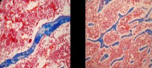 | Fig. 1- Sample of OC-joint - A: Bone trabeculae, failure of cartilage matrix mineralization - endochondral ossification (Masson’ trichrome, 40x); B: Trabecular bone, failure of cartilage matrix mineralization - endochondral ossification (Masson’s trichrome, 20x). |
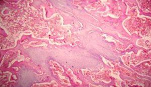 | Fig. 2 - Sample of OC-joint - Ephyfiseal plate, irregular cone-shaped, dilating cartilage canals, desorganization of chondrocytes columns and fracture areas (hematoxylin - eosin, 20x) |
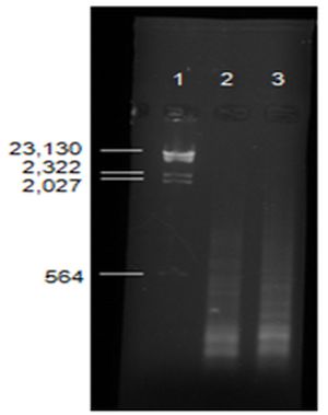 | Fig. 3- PD2 generated by RDA: Lane1 - λ hind marker; Lane 2 - PD2, OC-joint; Lane 3 - PD2, healthy joint. |
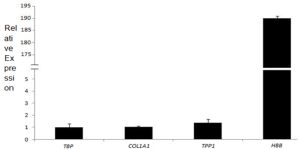 | Fig. 4- Validation of RDA results by RT-qPCR. The bar graph shows the collagen type I alpha 1 (COL1A1), tripeptidyl peptidase 1 (TPP1) and hemoglobin subunit beta (HBB) relative expression. The amount of mRNA measured in each sample was normalized using the Ct (threshold cycle) values obtained from the control gene TBP. The x-axis values represent the number of times the selected gene was expressed in the OC-joint compared to healthy joint. |
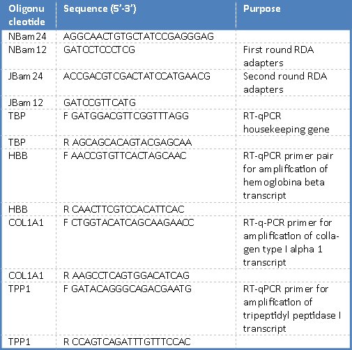 | Table 1- List of oligonucleotides and your aplications. |
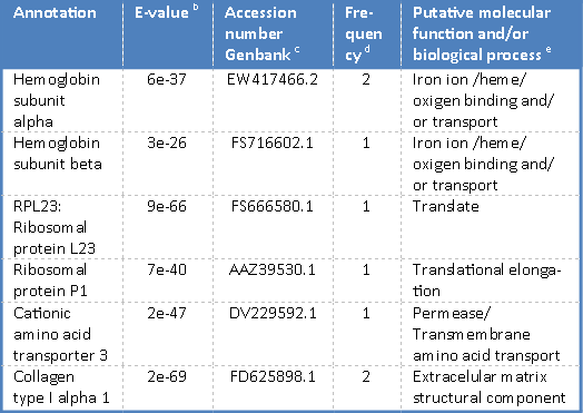 | Table 2- Summary of computational analysisa of genes obtained from forward RDA.library <sup>a</sup>Sequence quality was analysed by PHRED. Resulting sequences were compared to the GenBank database using BLASTx program. <sup>b</sup>E-value according to information from BLASTx searches of non-redundant database at NCBI. <sup>c</sup>Accession number of gene products in the GenBank database. <sup>d</sup>Frequency represents the number of clones found in a total of 11 high quality sequenced cDNAs clones. <sup>e</sup>Putative molecular function and/or biological process according to the Unigene, Interpro and Pfam classification system |
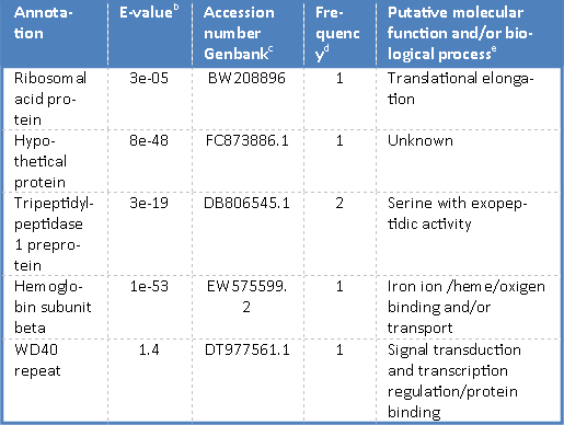 | Table 3 - Summary of computational analysisa of genes obtained from reverse RDA. library <sup>a</sup>Sequence quality was analysed by PHRED. Resulting sequences were compared to the GenBank database using BLASTx program. <sup>b</sup>E-value according to information from BLASTx searches of non-redundant database at NCBI. <sup>c</sup>Accession number of gene products in the GenBank database. <sup>d</sup>Frequency represents the number of clones found in a total of 11 high quality sequenced cDNAs clones. <sup>e</sup>Putative molecular function and/or biological process according to the Unigene, Interpro and Pfam classification system. |
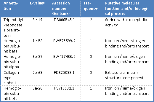 | Table 4- Sequences of forward and reverse libraries that had better quality and homology to genes in GenBank database <sup>a</sup>Sequence quality was analysed by PHRED. Resulting sequences were compared to the GenBank database using BLASTx program. <sup>b</sup>E-value according to information from BLASTx searches of non-redundant database at NCBI. <sup>c</sup>Accession number of gene products in the GenBank database. <sup>d</sup>Frequency represents the number of clones found in a total of 11 high quality sequenced cDNAs clones. <sup>e</sup>Putative molecular function and/or biological process according to the Unigene, Interpro and Pfam classification system. |