


ISSN : 0976-996X
EISSN : 0976-9978



LEONARDI L.1*, ROPERTO F.2, SFORNA M.3, ANGELI G.4, GIALLETTI R.5
1Università di Perugia Facoltà di Medicina Veterinaria Dipartimento di Scienze Biopatologiche e Igiene delle Produzioni Animali e Alimentari Via San Costanzo, 4 - 06126 Perugia – Italia
2Università di Napoli – Facoltà di Medicina Veterinaria – Dipartimento di Patologia e Sanità Animale – Via F. Delpino, 1 – 80137 Napoli – Italia
3Università di Perugia Facoltà di Medicina Veterinaria Dipartimento di Scienze Biopatologiche e Igiene delle Produzioni Animali e Alimentari Via San Costanzo, 4 - 06126 Perugia – Italia
4Università di Perugia – Dipartimento di Patologia, Diagnostica e Clinica Veterinaria – Via San Costanzo, 4 - 06126 Perugia – Italia
5Università di Perugia – Dipartimento di Patologia, Diagnostica e Clinica Veterinaria – Via San Costanzo, 4 - 06126 Perugia – Italia
* Corresponding Author : leonardo.leonardi@unipg.it
Received : 15-12-2011 Accepted : 16-01-2012 Published : 03-10-2012
Volume : 3 Issue : 1 Pages : 44 - 47
Vet Sci Res 3.1 (2012):44-47
DOI : http://dx.doi.org/10.9735/0976-996X.3.1.44-47
Conflict of Interest : None declared
Osteosarcoma is the most frequent malignant bone tumor in domestic animals and humans, representing 80-85% of malignant bone tumors in dogs [9, 10] and about 70-75% in cats. Only a few cases of osteosarcoma have been reported in horses with the majority in the mandible of young horses [3]. Maxillary osteosarcoma causes disruption of the bones with subsequent disruption of the dental arcade and interference with mastication [3, 5]. We describe a case of primitive parosteal fibrous maxillary osteosarcoma in a 16–year old Anglo-Arabian horse, hospitalized first for a clinical diagnosis of sinusitis. This case is also unusual in that generally maxillary fibrous osteosarcomas are low grade malignancies with minimal potential to metastatize, yet in this case the tumor had already spread to a regional lymph node by the time the horse was presented for examination, confirming the unpredictability of osteosarcoma.
Parosteal osteosarcoma, horse, maxilla, fibrous osteosarcoma, juxtacortical osteosarcoma.
Osteosarcoma is a malignant neoplasm of bone, in which malignant osteoblasts produce osteoid and immature bone [1] . The most characteristic feature of the osteosarcoma is the production of osteoid, which is nevertheless not always easily or conclusively identifiable, because it has no specific staining reaction. The tumor is higly malignant and usually metastatizes early. Osteosarcoma is the most common bone tumor in domestic animals and humans and it represent 80% of all malignant bone tumors in dogs [9,10] and 70% in cats [3] . Tumors of the head and neck are very rare in horses, but classic central osteosarcomas are still one of the most common sarcomas of the head and neck region [7] . The majority of reported cases of equine osteosarcomas occurred in the head and were more frequent in mandible of young horses [2,3,8,11] . The predominant subtype is fibroblastic osteosarcomas [3] . Many factors have been hypothesized to play a role in pathogenesis of osteosarcomas in humans and animals, including viruses, chemicals, ionizing radiations, chronic diseases, metallic implants, trauma, bone infarcts, certain skeletal diseases and disorders and other host factors such as body size and sex [13] . Pain and swelling are usually the initial clinical signs. Occasionally there is mild to moderate enlargement of the regional lymph nodes [4] Serological findings are not pathognomonic but increased alkaline phosphatase and lactic dehydrogenase (LDH) have been reported as unfavorable prognostic factors [4]
A 16 year old Anglo-Arabian horse presented to the Veterinary School of the University of Perugia with a large, firm painful mass of the head.
The horse, had bilateral purulent nasal discharge, normal shape and mobility of the nostrils, reduced air flow on the right, and inspiratory noise from the nostrils, and was hospitalized for sinusitis. The sinuses appeared misshapen, with right-sided asymmetry, and dullness on digital percussion of the right maxillary and frontal sinuses. Physical and endoscopic examination revealed complete occlusion of the right nostril due to the presence of a large, firm painful hard mass in the rostral portion of the right maxillary sinus overlying the palatal portion of the right maxillary alveolar ridge in the region of the molar teeth. Submaxillary lymph nodes were enlarged, and ultrasound revealed that one node had patchy hypo-and hyper-echogenic areas, and areas of mineralization. The radiographic examination showed the presence of a radiopaque mass, similar to bone, that was round with irregular edges and involved the maxillary sinus and to some extent the frontal sinus. Endoscopic examination confirmed a mass in the maxillary sinus which overflowed into the frontal sinus. Biopsy was attempted using a Jamshidi bone biopsy needle but the hardness of the mass did not allow a significant amount of tissue to be collected. A CAT scan was done to assess the boundaries of mass to determine if it could be surgically excised. CT findings reveal the presence of a neoformation developed expecially inside the caudal maxillary sinus, the frontal sinus and the concho-frontal sinus and characterized by a quite homogeneous bone-like density. Its borders was well distinguished from the normal skeletal base. There were no meaningful alteration of the bone of the skull except a deviation of the nasal septum and a osteolytic lesion of the maxillary bone were was performed a surgical opening of the maxillary sinus.
Gross examination revealed a hard mass 18.00 cm x 12.00 cm x 09.00 cm involving the right maxilla, the upper hard palate and the first and second molars. The tumor was ovoid and well demarcated by a thick pseudocapsule. Externally, the mass had multifocal areas of mineralization and necrosis and diffuse hemorrhages with peripheral reactive bone and focal reactive fibrous tissue. A complete necropsy was performed, and there was no gross evidence of metastasis in other organs or tissues.
Samples for histopathology were taken from several areas of the mass, fixed in 10% neutral buffered formalin, decalcified and embedded in paraffin. Sections of 4-5 μm were cut and stained with hematoxylin-eosin (HE). There were no areas in which the tumor penetrated into medullary bone of the maxilla. Histologic sections revealed that the tumor involved the superficial cortical bone of the maxilla and the upper hard palate. The tumor was characterized by ossified areas with well-developed trabeculae of woven bone partially embedded in a fibroblastic stroma. There was a moderate cellular atypia and moderate mitotic rate. The neoplastic cells were pleomorphic with milde signs of anisocytosis and anisokariosis. The intertrabecular stromal cells were composed of a mixture of both spindle-shaped and round cells. In addition, there were islands of lymphocytes, osseous necrosis, hemorrhage and rare multinucleated cells compatible with osteoclasts. A submaxillary lymph node contained the same neoplastic cells described in the primary mass, which also involved related vessels. The findings were most consistent with a medium-grade fibroblastic osteosarcoma with irregular islands of osteoid surrounded by pleomorphic sarcoma cells. The diagnosis was confirmed by 2 other pathologists who reviewed the original histopathological samples. The final diagnosis was medium-grade parosteal fibrous maxillary osteosarcoma with metastasis to a regional lymph node.
Immunohistochemistry (IHC) was performed using the avidin-biotin complex method (ABC). The following primary antibodies were used: pancytokeratin (1:200, pH 9.0, Dako, Denmark, AE-1/AE-3) and polyclonal rabbit anti-S100 (1:1000; Dako, Denmark). Three-amino-9-ethylcarbazole (AEC Substrate-Chromogen, Dako, Denmark) was used as a chromogen and Carazzi’s hematoxylin as a counterstain. Negative controls were performed in the same manner, omitting the primary antibody. The malignant cells were negative for both pancytokeratin and S-100.
Parosteal osteosarcoma was first descripted in the long bones of humans in 1950 by Geschickter and Copeland [7] and craniofacial parosteal osteosarcoma was first reported in humans in 1961. To the authors’ knowledge, this case represent the first report of a parosteal fibrous maxillary osteosarcoma in a horse. Although osteosarcoma is the most frequent malignant bone tumor in humans and animals, this tumor is very rare in horses, particularly in the maxilla. Radiographic investigations demonstrated continuity with cortical bone in contrast to periosteal lesions, which are generally distinguished by an intact cortex [3,14] In humans parosteal osteosarcomas usually overgrowth their base of origin, whereas the periosteal variant tends to remain within the confines of its base [6,7,12] . This case is unusual in that generally maxillary fibrous osteosarcomas are low grade malignancies with minimal potential to metastasize, yet in this case the tumor had already spread to a regional lymph node by the time the horse was presented for examination, confirming the unpredictability of osteosarcoma. An accurate diagnosis requires correlation of clinical, radiographic, histologic and immunohistochemical data. The horse was euthanized at the time of diagnosis.
[1] Adler C.P. (2000) Bone diseases, macroscopic, histological and radiological diagnosis of structural changes in the skeleton.
» CrossRef » Google Scholar » PubMed » DOAJ » CAS » Scopus
[2] Barber S.M., Clarck E.G., Fretz P.B. (1983) Journal of American Veterinary Medical Association, 182: 700-702.
» CrossRef » Google Scholar » PubMed » DOAJ » CAS » Scopus
[3] Bush J.M., Fredrickson R.L., Ehrart E.J. (2007) Equine Osteosarcoma: a series of 8 cases. Veterinary Pathology, 44: 247-249.
» CrossRef » Google Scholar » PubMed » DOAJ » CAS » Scopus
[4] Campanacci M. (1999) 2nd Ed. 1999, Springer-Verlag Wien New York.
» CrossRef » Google Scholar » PubMed » DOAJ » CAS » Scopus
[5] Daffner R., Fox K.R., (2002) Galey K.R. Fibroblastic osteosarcoma of the mandible. Skeletal Radiology, 31: 107-111.
» CrossRef » Google Scholar » PubMed » DOAJ » CAS » Scopus
[6] Desai D., Pandith S., Jeergal P.A., Arathi K., Saini R. (2010) Fibroblastic variant of osteosarcoma: a challenge in diagnosis & management. The Open Dentistry Journal, 4 211-217.
» CrossRef » Google Scholar » PubMed » DOAJ » CAS » Scopus
[7] Hewitt K.M., Ellis G., Wiggins R., Bentz G. (2008) Parosteal osteosarcoma: case report and review of the literature. Head and Neck, 122-126.
» CrossRef » Google Scholar » PubMed » DOAJ » CAS » Scopus
[8] Jenner F., Solano M., Gliatto J., Lavalles S., Kirkerhead C. (2003) Equine Veterinary Journal, 35, 214-216.
» CrossRef » Google Scholar » PubMed » DOAJ » CAS » Scopus
[9] Leonardi L. (2001) Indagine retrospettiva sulla frequenza dell’osteosarcoma in campioni bioptici e chirurgici. Atti A.I.P.Vet, 27-28 maggio, Messina, Italy.
» CrossRef » Google Scholar » PubMed » DOAJ » CAS » Scopus
[10] Leonardi L., Brachelente C., Sforna M., Capuccini S., Mechelli L. (2005) 23rd Meeting of European Society of Veterinary Pathology, 07-10, Naples – Italy.
» CrossRef » Google Scholar » PubMed » DOAJ » CAS » Scopus
[11] Livesey M.A., Wilkie I.W. (1986) Equine Veterinary Journal, 18, 407-410.
» CrossRef » Google Scholar » PubMed » DOAJ » CAS » Scopus
[12] Mc Guff S., Heim-Hall J., Holsinger C., Jones A.A., O’Dell D.S., Hafemeister A.C. (2008) Journal of American Dent. Association, 139, 1052-1059.
» CrossRef » Google Scholar » PubMed » DOAJ » CAS » Scopus
[13] Meuten D.J. (2002) Tumors in Domestic Animals, 4th ed, 266-267.
» CrossRef » Google Scholar » PubMed » DOAJ » CAS » Scopus
[14] Zaruby J.F., Williams J.W., Lovering S. (1983) Canadian Veterinary Journal, 742-744.
» CrossRef » Google Scholar » PubMed » DOAJ » CAS » Scopus
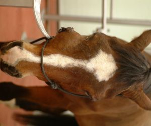 | Fig. 1- ulcerated mass in the right maxilla before surgery. |
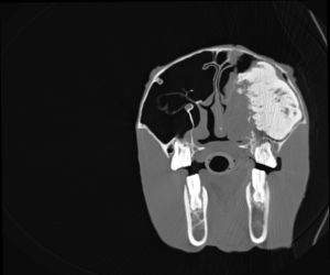 | Fig. 2- CT findings reveal the presence of a mass involved the caudal maxillary sinus, the frontal sinus and the concho-frontal sinus, characterized by a quite omogeneous bone-like density. |
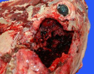 | Fig. 3- gross features of the tumor with destructive growth inside the maxillary involved bone. |
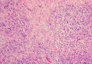 | Fig. 4- fibroblastic pattern, the tumor is composed of sheets of streaming neoplastic spindloid cells, with sheets of osteoid surrounded by sarcomatous cells with large and irregular nuclei. h&e stain, 10x. |
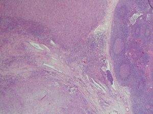 | Fig. 5- submaxillary lymph node: direct tumor infiltration with endolymphatic microfoci of tumor. h&estain, 10x. |
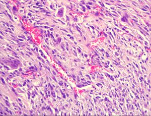 | Fig. 6- fibroblastic pattern with activation of osteoclastic activity and osteoid production surrounded by pleomorphic sarcoma cells and mitotic figures. h&estain, 20x. |
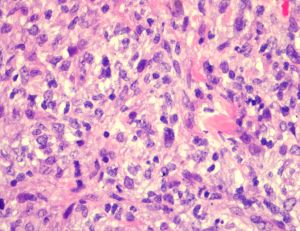 | Fig. 7- fibroblastic pattern with activation of osteoclastic activity and osteoid production surrounded by pleomorphic sarcoma cells and mitotic figures. h&estain, 40x. |