


ISSN : 0976-8920
EISSN : 0976-8939



BUSHRA AQUIL1, ARIF TASLEEM JAN2, NEERA BHALLA SARIN3, QAZI MOHD. RIZWANUL HAQ4*
1Department of Biosciences, Jamia Millia Islamia, New Delhi-11002, India.
2Department of Biosciences, Jamia Millia Islamia, New Delhi-11002, India.
3School of Life Sciences, Jawaharlal Nehru University, New Delhi-110067, India.
4Department of Biosciences, Jamia Millia Islamia, New Delhi-11002, India.
* Corresponding Author : qmrhaque@gmail.com
Received : 04-04-2012 Accepted : 07-05-2012 Published : 01-06-2012
Volume : 3 Issue : 2 Pages : 64 - 77
J Crop Sci 3.2 (2012):64-77
DOI : http://dx.doi.org/10.9735/0976-8920.3.2.64-77
Conflict of Interest : None declared
Banana, fourth most important food commodity on earth, has great socio-economic significance in India. As a staple food, it contributes to food security of millions of people in developing world. Despite playing a key role in the economy of many developing countries, its production and as such export is constrained by many biotic as well as abiotic factors. Increase in production through minimization of yield loss has been emphasized as one of the thrust area and biotechnological advances such as genetic transformation, is playing a key role to handle the situation. Besides protecting banana against wide range of diseases, genetically modified bananas have also been advocated as carrier for vaccines and as a source of carotenoids that can counteract debilitating vitamin A deficiency. In order to augment micropropagation of banana through different explants and different types of organogenesis and to avoid the constraints imposed by pests and pathogens, transgenic approaches using particle bombardment or Agrobacterium-mediated transformation using different transgenes is preferentially considered for the improvement of banana crop. This article deals with studies on micropropagation and genetic transformation that are preferably used for the enhancement of banana production though protection against biotic and abiotic stresses.
Banana, Biotic and abiotic stress, Genetic transformation, Micropropagation.
Banana is the common name of genus Musa sp., having great importance in the world due to their commercial importance and high nutritional value. Besides, being a good source of proteins and carbohydrates, it is considered as a source of vitamins (Vit A, C, E, K, B1, B2, B3, B6 and vitamin B9), minerals such as potassium, sodium, calcium and magnesium along with trace amounts of iron, zinc and carotenoids [1] . Banana has also been found effective against colorectal cancer [2] , breast cancer [3] , and renal cell carcinoma [4] . Most, but not all banana cultivars are triploid (2n = 3x = 33 chromosomes), with AAA, AAB and ABB genome constitution. These triploid genotypes are virtually or completely sterile and develop their fruit by vegetative parthenocarpy. They are derived from inter- and intraspecific hybridisation between two different species of genus Musa; M. acuminata and M. balbisiana, designated with the diploid genomes AA or BB respectively [5] . Among Eumusa species of banana, Musa acuminata is the most widespread, with Malaysia [6] or Indonesia (Nasution [7,8] as its centre of diversity.
Banana ranks fourth (in terms of production) in the world and is second most significant fruit crop in India next to mango [9] . Presently, banana is grown in around 150 countries across the world on an area of 10 million ha with a total production of 100 million tons, out of this 43% is contributed by dessert banana type, while as cooking banana makes up approximately 57% [10] . Current world dessert banana production is approximately 67 million tons per year [10] , but only 20% of it enters the world trade. Major dessert banana-producing countries are India, Philippines and Brazil, but neither of these exports significant quantities [Table-1] . By comparison Brazil, the fifth largest producer, exports approximately 67% of its bananas and is as such the largest contributor of dessert bananas to world trade. Dessert cultivars of banana and plantain (a subgroup of cooking banana) banana are seedless. Their propagation of progeny generally takes place by planting the suckers. India, with an annual production of 26.22 million tonnes from 404,000 ha area under cultivation, is the largest producer of banana. It contributes 27% to world production and about 37% of t total fruit crop production in the country [10] . Among the Indian states, Maharashtra that contributes maximum area of about 90,000 ha for banana cultivation, accounts for 25% of the total banana production in India followed by Tamil Nadu (20%), Gujarat (15%), Karnataka (10%) and Andhra Pradesh (10%). Despite being most productive (Jalgaon in Maharashtra and Trichy in Tamil Nadu), their contribution accounts only 0.1% to World trade.
Constraitns As with all other crop species, banana production faces major challenges from biotic as well as abiotic stresses.
Banana production is limited due to several diseases and pests, such as fusarium wilt (Fusarium oxysporum f. sp. Cubense), black sigatoka (Mycospharella fijiensisi), banana xanthomonas wilt (Xanthomonas campestris pv. musacearum), viruses (Banana bunchy-top virus, genus Nanovirus and Banana streak virus, genus Badnavirus), weevils, and nematodes [11] .
Panama disease or Fusarium wilt, caused by the fungus Fusarium oxysporum, has devastated banana production and is widely regarded as one of the most destructive plant diseases [12] . Once established in an area, Fusarium cannot be controlled chemically by fungicides or solid fumigants, or by cultural practices such as rotations or soil amendment, so the only long-term option for continuing banana production is replacement of a susceptible variety with a resistant variety [13,14] . However, most commercial varieties are susceptible to ‘Tropical Race 4’ [15] . Although a number of varieties have been identified with resistance genes that may be useful in breeding or gene-transfer programs, these varieties have weaknesses that makes them unsuitable as replacements for ‘Cavendish’ [14] . Another fungal disease, Black sigatoka leaf spot or black leaf streak disease (BLSD, Mycosphaerella fijiensis) has been serious, with infection leading to around 50% crop losses [16] There is some genetic resistance in Musa with potential for exploitation [17] , and genomic studies of the pathogen (BLSD, Mycosphaerella fijiensis), including complete sequencing, are underway [18] .
A bacterial wilt caused by Xanthomonas is spreading rapidly in East Africa; although control of disease spread by cultural practices is being attempted, a long-term solution may again come through the genetic resistance.
Viral diseases of banana include various diseases such as Banana Bunchy Top and Banana Streak caused by BBTV and BSV respectively are controlled mainly by eradication of infected plants. Harper et al. (1999) showed the BSV-related sequences are integrated within the nuclear genome although integration is not an essential part of the viral life cycle [19] . Hull et al. (2000) and others have speculated that the presence of integrated copies may confer virus resistance through induction of transcriptional or post transcriptional gene silencing of homologous sequences, and since then it has become clear that expression of these elements give rise to a strong viral infection [20,21] .
Burrowing nematodes (Radopholus similis and Pratylenchus spp.) and weevil (Cosmopolites sordidus) pests also constrain banana production, with little genetic resistance in widely grown cultivars. Their infection often leads to conditions where plantations becoming uneconomic and being abandoned. No known source of desired level of resistance exists within the banana/plantain gene pool [22-24] .
Abiotic stresses caused by shortage or excess of water, salinity, wind or temperature, affects the crop yield [5] . Plants can tolerate short periods of drought because of their water-filled reserves [25] . Lack of water is associated with ‘maturity bronzing’ effect, manifested by discoloration of mature bananas and cracking of the skin [25] . A soil pH in the range 5.5-7.5 is suitable for growing bananas, with pH 5.5 considered as optimal [26] . A low pH however solubilizes elements like iron, aluminium and manganese; these can be toxic and have negative effects on the plant growth. A low pH also reduces the availability of other nutrients such as calcium and higher than 6.5, can reduce the availability of trace elements such as boron, zinc, copper and iron [26] .
Despite high water requirements, water logging of the soil often results in oxygen starvation of the roots [27] . Oxygen deficiency for more than 6 hours results in root tip death, which in turn leads to branching of the roots [28] When sufficient water becomes available and roots recommence growing, it may result in multiple branching, giving it appearance of ‘witches broom’ [28] . Macronutrients required by banana plants include nitrogen, potassium, phosphorus, calcium, magnesium and sulphur. Deficiency of potassium results in reduced buoyancy, which interferes with the post harvest production processes; the fruit sinks when dipped in hot water for the treatment against certain diseases [29] . The micronutrients required by bananas include boron, iron, manganese, copper, zinc, molybdenum, chlorine and cobalt. Deficiencies in these elements lead to morphological malformation of the leaves, reduced growth and yield and poor fruit quality [25] . Boron deficiency alone can result in fruit that does not ‘fill’ [26] Bananas do not thrive in areas of high salinity, although some varieties show tolerance than others. High levels of sodium result in reduced crop growth due to a reduction in osmotic pressure of the soil, which leads to an increase in ions that are toxic to the plant [30-32] .
All Musa species grow best in the open sun provided that the moisture is not limiting [6] Deep shade causes stools to die [6,25] . Fire generally does not destroy the banana plant; they recover by regrowing from the corm [25] High humidity, >95%, during the final stages of ripening can lead to ‘splitting’ of the fingers [25] . Bananas are also susceptible to strong winds, which can twist and distort the crown. The leaves can also be shredded by winds thus interfering with metabolism [33] . Low temperatures retard growth although susceptibility to cold varies among cultivars [26] . Impact of cold stress on plant growth includes; non-emergence of bud from the stem at flowering time, cessation of root growth at temperatures below 130C and destruction of the plant by frost (although the corm normally remains viable) [26] . Choke Throat occurs when the bunch gets mapped in the pseudostem at various stages of emergence. Less severe cases result in bunches that only partially emerge from the pseudostem and are thus susceptible to disease because they are difficult to cover [34] .
Keeping in mind the above stresses, it is emphasized that an ideal ideotype cultivar is one which is disease and pest resistant, high yielding, photosynthetically efficient, early maturing, display minimum delay between consecutive harvests, short stature, strong roots for optimal nutrient uptake and greater resistance to wind damage. Considering the global importance of banana, there is a great potential to improve disease-free and high-yielding cultivars. As a step towards this, development of efficient organogenesis and genetic manipulation techniques will come up with new opportunity for the genetic improvement of banana.
In vitro organized and/or unorganized growth of new plantlets is called micropropagation. Since germination frequency of seed is extremely low, embryo culture is preferred for classical breeding experiments [35] . Major applications of shoot tip culture are mass clonal propagation and germplasm conservation. In mass clonal propagation, existing shoot tips are stimulated to multiply rapidly, while as in germplasm conservation, multiplication rate is slowed down. On induction, embryogenic callus from suitable explants in semi-solid medium containing high auxin concentration, it is transferred to liquid medium where it gives rise to embryogenic cell suspension (ECS). As such suspension possesses high regeneration capacity for mass clonal propagation, they are considered as main source for regenerable protoplasts in banana [36] . More importantly, they are preferred target material for induced mutations and genetic engineering. In vitro banana tissue culture plantlets that show uniform and vigorous growth with have a shorter harvesting period, have higher survival rate than suckers in natural surroundings. Commercial application of banana by in vitro culture usually involves shoot multiplication [37] . This technique can increase the rate of seedling production and improve the seedling quality such as uniformity and being true to parental type.
Regeneration through organogenesis.
Shoot tips being easy to culture are extensively preferred as starting material for micropropagation for a wide range of banana cultivars [38-41] . Ganapathi et al. (1998) reported regeneration of banana, Lal Kela (AAA genotype) from shoot tips and obtained 5-6 shoots per explants [42] . Priyono (2001) reported that micropropagation of Musa paradisiaca through cormlet initiation by in vitro culture of apical meristem slices [43] . Josekutty et al. (2003) established the efficient micropropagation of Apat regular and Apat fissuse (cooking bananas) using shoot meristem [44] . Hamide and Pekmeze (2004) used shoot tips to multiply banana cultivars dwarf Cavendish [45] . Diro and Staden (2005) also reported rapid in vitro protocol for multiplication of Enset vetricosum from shoot tips [46] . Kanchanapoom and korapatchaikul (2012) reported induction of yellow compact calluses from in vitro-grown shoot tips of diploid bananas (Musa acuminata, AA group) ‘Kluai Sa’ and ‘Kluai Leb Mu Nang’ [47] . Unlike other methods that need for field access and seasonal dependence, main advantage of this procedure is that it bypasses this procedure.
For microprogation of banana, use of leaf sheath and corm has the advantage that it can be applied to many banana varieties irrespective of the genotype. Okole and Schulz (1996) regenerated banana cultivar ‘Williams’ (AAA genotype) and two plantain cultivars ‘Horn’ (AAB) and ‘Cachaco’ (ABB) from leaf bases and corm slices [48] . Venkatachalam, et al. (2006) reported direct shoot and cormlet regeneration from leaf explants of ‘silk’ banana [49] .
Bakry et al. (1985) gave the first report on vegetative shoots from in vitro culture of inflorescence of banana [50] . Cronauer and Krikorian (1988) reported adventitious shoot bud formation from the determinate floral buds in plantain (AAB) [51] . According to them the apical dome has a role to play in re-differentiation process. However, Doreswamy and Sahijram (1989) who achieved micropropagation of Cavendish cultivar ‘Rasthali’ (AAA), reported that the apical dome per se did not participate in re-differentiation process and, therefore, did not directly contribute to development of vegetative shoots [52] . Balakrishnamurthy and Rangaswamy (1988) reported in vitro shoot regeneration from floral apices of banana cultivars, ‘Robusta’ (AAA) and ‘Monthan’ (ABB) [53] . It is believed that different hormones play a role in the reversal of vegetative stage from the reproductive stage of banana. According to Ling et al. (1990), addition of 2, 4-D; 2, 4, 5-T or kinetin to MS medium could induce the reversion of the reproductive stage to vegetative stage in ABB group of banana [54] . Murali and Duncan (1991) induced vegetative stage in Gros Michel banana flower bud, when cultured on MS medium supplemented with BAP (5mg/l) and IBA (1mg/l) [55] . Chinsuk and Silayoi (2001) obtained 5.71 shoots/explants in MS medium supplemented with 7mg/l BAP [56] . According to Cirad (2003) immature male flowers are the most successful explants for somatic embryogenesis [57] ; however, a rapid decline in embryogenic response soon after harvest as well as seasonal dependence makes it necessary that cultures must be developed quickly from harvested flowers [58] .
Okole and Schultz (1996) studied the micro-cross sections of banana cultivar ‘Williams’ (AAA) and two plantain cultivars ‘Horn’ plantain (AAB) and Cachao (ABB) for adventitious shoot and callus formation [48] . The micro-cross sections were derived from the lower to upper leaf segments of all three genotypes. An average of 15 shoots buds were obtained from micro-cross sections derived from each comb. The shoot buds could be successfully regenerated to give rise to plantlets.
The nature and physical state of the culture medium plays an important role in in vitro culture of Musa species. Semi solid medium is widely used for routine multiplication of bananas all over the world, although use of liquid medium for in vitro micropropagation is often described as a way of reducing cost of producing plantlets. Sterilization of the medium can be performed by ultra-filtration i.e. filtration of the solution through filters of pore size less than 0.45 m, rather than autoclaving. Despite the advantages of shorter sub culturing time for explants, use of liquid medium involves the problem of asphyxia of explants as a result of immersion. The most commonly used preventive methods are based on the principle of partial immersion of explants, by using Filter Paper Bridge or cellulose, Rockwool etc. [59] . Bhagyalakshmi and Singh (1995) compared static liquid, agitated liquid and agar-gelled medium for shoot tip multiplication and plant survival in three cultivars of banana i.e. Cavendish, Bluggoe and Silk using shoot tip explants and reported maximum number of shoots as well as maximum survival ex vitro in static liquid medium coupled with judicious selection of shoot cultures and a brief exposure of the same in gelled medium without BAP [60] . Chinsuk and Silayoi (2001) reported shoot formation from inflorescences of banana cv. Kluai Khai (Musa acuminata ‘Klaui Khai’) by sub-culturing in liquid MS medium [56] . Temporary immersion system (TIS) is a simple method that is used to multiply in liquid medium, organogenic cultures such as nodules, meristem clumps or compact shoot clusters, somatic embryos and cells, and for growth of bulbs, corms or microtubers [61] . This system has been successfully used in the micropropagation of bananas of the AAA [62,63] and AAAB groups [64] . Matsumoto and Brandao (2002) compared TIS and permanent immersion system (Explants permanently dipped in liquid medium and sterilized filter air injected in medium every four hours for five seconds) with conventional culture system using shoot tip explants of cultivar Maca (genotype AAB), and observed that TIS and permanent immersions system respectively produced 3.7 and 12 times more plant material than the conventional culture system [65] .
Proliferation rate of shoot and elongation are affected by the type and concentration of plant growth regulators. Cytokinins and auxins are used as growth regulators for in vitro propagation of Musa spp. As concentration of exogenous cytokinin appears to be the main factor affecting shoot multiplication, most widely used and most effective cytokinin for this purpose is adenine based cytokinin; N6-benzylaminopurine (BAP) [38,40,45,66] . Others include isopentyladenine (2-ip) [38] , zeatin [67] and kinetin [68] .
Cronauer and Krikorian (1984a) obtained 9.1 shoots per explants during in vitro multiplication of Phillippine lacatan and Grand naine, on a modified MS medium supplemented with 10 M BAP [38] while as Rahman et al. (2002) obtained 4.52 shoots per explants on the same media in variety Bari-1. It indicates different genotypic response towards the cytokinin BAP [69] . Noor Aziah and Khalid (2002) used higher concentration of BAP for regeneration, using whole meristems and scalps as explants [70] . Scalps were induced on MS medium supplemented with coconut water and BAP (75
M). The average number of shoots produced from scalps was six times more than that produced from a single shoot tip. Venkatachalam et al. (2006) achieved direct regeneration from leaf sheaths of silk banana (AAB) on MS medium supplemented with BAP (22.4
M) [49] . Thiadiazuran (TDZ) is a urea based cytokinin, which is frequently used in banana micropropagation. Hamide and Pekmezc (2004) tested the effects of BAP (5, 10, 20 and 30  M) and TDZ (0.4, 1, 2 and 3
M), either alone or in combination with 1
M indole acetic acid (IAA) for shoot multiplication in three banana types [45] They observed that in all the three type, shoot proliferation and elongation were significantly greater with TDZ as compared with BAP. Also each cytokinin, in combination with 1
M IAA increased shoot proliferation and elongation more than when used alone. Strosse et al. (2008) cultured shoot tip explants to determine the influence of five cytokinins [BA] kinetin, isopentenyladenine (2iP), zeatin, and thidiazuron (TDZ)] each at three concentrations (1, 10 and 100
M) added to the basal corn shoot multiplication (CSM) medium, on multiple shoot formation [71] . When shoot tips of banana variety Williams (AAA) were cultured on basal CSM medium devoid of plant growth regulators, all explants grew into elongated single shoots. The highest number of explants developed into elongated shoots. The highest number of explants developing into multiple shoots was observed with TDZ (up to 100%) followed by BAP (up to 92%). These studies suggest that a combination of cytokinins, with or without auxin have been used for enhancing shoot proliferation in banana regeneration.
Growth retardants have been reported to be used along with cytokinins to increase the number of multiple shoots per explants. Albany et al. (2002) introduced growth retardants ancymidol (ANC), paclobutrazol (PBZ) and diaminozide (DAM) respectively, in liquid cultures constituting temporary immersion system in order to reduce the size of the shoots to allow better use of space inside the culture vessel and increase their numbers [72] . In liquid shake cultures ANC and PBZ (irrespective of concentration), promoted bud formation in clusters, but with a reduced size and compact shape. It took five successive subcultures in ANC or PBZ (2.5 mg/l) containing medium to recover the normal morphology of the shoots as compared to those grown in hormone-free medium without growth retardants. However, during the acclimatization stage, plants multiplied in PBZ and DAM containing media or plants multiplied in ANC containing media showed reduced height in comparison to control plants.
Plant embryogenesis is the process that produces a plant embryo from a fertilized ovule by asymmetrical cell division and differentiation of undifferentiated cells into tissues and organ. Somatic embryogenesis involves the development of embryos from somatic cells. The bipolar structure of somatic embryo contains both shoot and root meristems. This in vitro morphogenetic pattern is a multi-step regeneration process, starting with the induction of pro-embryogenic masses, followed by somatic embryo formation, maturation, desiccation and plant regeneration, in response to physical and chemical treatments [73] . In bananas and plantains, four procedures exist for the development of embryogenic cell suspensions (ECS). They differ mainly in the source of the explants: zygotic embryos [51,74] , rhizome slices and leaf sheaths [75] , immature female flowers [76,77] and multiple meristem cultures [78,79] . Since most edible banana cultivars rarely set seed, zygotic embryos are of limited value as starting material. On the other hand, the scalp methodology relies on proliferating meristem cultures as explants. This involves an extensive material preparation phase preceding induction of embryogenesis. In contrast, the starting material for the widely used male-flower technology can be collected directly from the flowering banana plants. However, factors such as fast decline of the male flower embryogenic response soon after harvest; seasonal dependence; direct field access and inoculation of explants quickly after harvest limits this methodology [58] . Besides that, this method cannot be applied to varieties such as False Horn plantains and Harton plantains, which do not produce make flowers.
Zygotic embryos
Cronauer and Krikorian (1988) reported somatic embryogenesis in a seeded diploid ornamental banana Musa ornata Roxb [51] . The embryos were excised from the seeds and placed on semi-solid MS medium supplemented with different concentrations of 2, 4-D (0.5, 1, 2 mgl-1) and 5% coconut water. Embryo germination was obtained on SH [80] salts followed by transfer to MS basal medium. Plantlets were obtained using filter paper bridges in liquid half-strength SH medium supplemented with 1% sucrose. Escalant and Teisson (1989) obtained callus from immature zygotic embryos of Musa acuminata (AA) and Musa balbisiana (BB) [74] . After removing seed coat from the seeds, embryos were separated from albumin. These embryos were cultured in M1 medium [MS salts in which macroelemetns were diluted two-fold but supplemented with 1.47 mM KH2PO4 and vitamins of supplemented with 2.2 M BAP [81] . After one month of culture, 80% of these embryos germinated and formed shoots. White compact heterogeneous callus appeared at the base of about 20% of these shoots. These callus were isolated and cultured on M2 medium (same as M1 but macro-elements were not diluted) supplemented with 9
M dicamba or 7.5
M picloram under darkness. After one month somatic embryos appeared on the compact part of embryo. These somatic embryos converted to plantlets on M2 medium supplemented with 5.3
M NAA. In another study. Kanchanapoom and korapatchaikul (2012) reported Yellow compact calluses were induced from in vitro-grown shoot tips of diploid bananas (Musa acuminata, AA group) ‘Kluai Sa’ and ‘Kluai Leb Mu Nang’ on a modified Murashige and Skoog (MS) medium containing 100 mg/L malt extract, 50 mg/L proline, 50 mg/L cysteine, 100 mg/L glutamine, 1 mg/L biotin, 7 mg/L Dicamba and 2 mg/L TDZ [47] . Green shoot buds were induced after transfer of the yellow compact calluses to the same MS medium, but supplemented with 1 mg/L NAA and 3 mg/L BA so as to achieve plant regeneration through organogenesis in callus cultures.
Novak et al. (1989) reported embryogenic callus formation by inoculating leaf bases and rhizome tissue explants of triploid cooking bananas, Cardaba (ABB genome), SH-3362 (AA genome) and Bocadillo (AA genome), in modified SH medium supplemented with an auxin, dicamba (3,6-dichloro-2-methoxybenzoic acid) (20 M) and a cytokinins, TDZ (5
M). Cell suspension cultures after 3-4 weeks inoculation in medium supplemented with zeatin (5
M) developed somatic embryos [75] .
Ma (1991), Escalant et al. (1993) and Jalil et al. (2003) obtained embryogenic cell suspension (ECS) cultures and plant regeneration of Musa acuminata by inoculating male flowers on MS medium supplemented with 1 mgl-1 biotin, 100 mgl-1 malt extract, 100 mgl-1 glutamine, 18 M 2,4-D, 5.4
M IAA, 5.7
M NAA, 30 gl-1 sucrose and gelled with 2.6 gl-1 BAP (M4 medium) [82-84] Grapin et al. (1996) and Shii et al. (1992) also reported plant regeneration from embryogenic calli obtained by subculturing male flowers on MS medium [85,86] . Ganapathi et al. (1998) established embryogenic cultures using young male flowers in five cultivars of banana, namely Rasthali (AAB), Basrai (AAA), Shreemanti (AAA), Lokhandi (AAA) and Trikoni (AAA) [42] . Embryogenic callus continued to rapidly proliferate on MS medium supplemented with 0.22
M BA and 1.14
M indole-3-acetic acid (IAA) and somatic embryos developed on the same medium. Green plumules emerged from the embryos followed by development of roots within a span of 6 to 8 weeks upon transfer to
strength MS medium supplemented with 0.5 g/l malt extract and 0.1% activated charcoal. Subsequently normal plant development was obtained following transplantation into paper cups in the greenhouse.
Grapin et al. (2000) reported banana regeneration via, cell suspension of false Horn plantain (Musa AAB) using female flower as explant [77] . Gomez et al. (2000) regenerated plantlets of hybrid cultivar FHIA-18 (AAAB) through formation of somatic embryos in liquid medium by using male flowers [87] . Perez et al. (2005) reported new methodology for establishing ECS and embryo germination in banana (Musa sp. AAA, ‘Grand Naine’) from immature floral buds of the male inflorescence [88] . They used two different culture Media to establish cell suspension cultures; one MS salts supplemented with 4mg/l of 2, 4-D and other medium having 1 mg/l of 2, 4-D. Floral meristems were inoculated separately in 50 ml Erlenmeyer flasks with 5 ml of respective culture medium. The flasks were kept in an oribital shaker at 100 rpm in darkness. The ECS were obtained in 100 days in both culture media. In both media cell suspension cultures comprising of small and irregular cell aggregates were formed. Sedimented cells (0.5 ml) were plated on semisolid medium for regeneration of plantlets, where 78% of the embryos germinated after 35 days of culture.
Secondary somatic embryogenesis (SE2) is the process of induction of new somatic embryos from pre-existing embryos [89] . Khalil et al. (2002) used immature male flower buds to regenerate banana cultivar Dwarf Brazilian (Musa sp. AAB) through formation of SE2 [90] . Primary somatic embryos were produced when explants of immature male flower buds were cultured on MS medium supplemented with 1 mg/l biotin, 100 mg/l malt extract, 100 mg/l glutamine, 4 mg-1 2, 4-D, 1 mgl-1 IAA, 1 mgl-1 NAA, 30 gl-1 sucrose and 2.6 gl-1 Phytagel (M1 medium) and then transferred to M1 medium supplemented with 200 mgl-1 casein hydrolysate and 2 mgl-1 proline. Suspension cultures were initiated from embryogenic tissues placed in liquid medium supplemented with 2, 4-D (1 mgl-1), biotin (1 mgl-1), L-glutamate (100 mgl-1), malt extract (100 mgl-1), and sucrose (45 gl-1). The somatic embryos developed when suspension culture was aspirated on MS medium supplemented with biotin (1 mgl-1), kinetin (0.5 mgl-1) zeatin (0.2 mgl-1), sucrose (45 gl-1), and phytagel (2.6 gl-1). Differentiated embryos were transferred to MS medium supplemented with 5 mgl-1 BAP for maturation and cultured on hormone-free MS medium for germination and development into plantlets. Approximately 90% of the somatic embryos germinated and developed into plantlets and 900-1050 plantlets were obtained from initial starting material amounting to 90% regeneration of 0.5 ml packed cell volume (PCV) of the suspension culture in 4-5 months. Wei et al. (2005) established ECS from male-flowers of Musa accuminata cv. Mas (AA), a major banana variety of the South East region [91] . Sidha et al. (2007) tested immature male flowers (IMFs) of different banana cultivars (Ardhapuri, Basrai, Grande Naine, Lalkela, Mutheli and Shrimanti) for callus induction on medium containing 2,4-D, IAA and NAA [92] . In terms of callus inducting from IMFs of AAA genomic group, Lalkela showed highest response (77.7%) followed by medium response in Shrimanti (52.2%), Basrai (51%), Grande Naine (42.5%), Ardhapuri (42%) and Mutheli (40%). Compared to this group, Safed Velchi (BB) was found to be highly responsive to 1MF culture (70%) similar to Lalkela. Embryogenic response was the highest in Lalkela (83.3%), followed by Grandenaine (62%) Ardhapuri (50%), Basrai and Mutheli (45%) and Shrimanti (40%) while, Safed Velchi showed less response (20%). Experiments with different auxins (2,4-D & its analogs, dicamba, Picloram and Phenyl acetic acid) showed varied response among the cultivars tested.
Perez and Rossel (2008) obtained induction of somatic embryogenesis and suspension-derived plant regeneration of banana (Musa AAA, cv. ‘Dwarf Cavendish’) from inflorescences of male flowers [93] . Ghosh et al. (2009) developed an efficient method for somatic embryogenesis and plant regeneration of Cavendish banana cultivar Robusta (AAA) [94] . The ECS was established using immature male flowers and percentage of embryogenic calli and distinct globular embryos obtained was 10.3 and 11.1, respectively. Approximately 48.67% of the cultures showed the appearance of green nodular callus from the base of the explants within 3-4 months of culture in MS medium supplemented with 2,4-D (4 mgl-1), IAA (1 mgl-1), NAA (1 mgl-1), biotin (1 mgl-1) and 3% sucrose. At this stage the responding cultures were transferred to semisolid MS medium supplemented with 2, 4-D (1 mgml-1), biotin (1 mgml-1), malt extract (100 mgl-1), glutamine (100 mgml-1), and 4.5% sucrose (M2 medium), in which about 10.3% of the transferred cultures formed white friable embryogenic calli. Distinct globular embryos were observed in 11.1% of embryogenic cultures on M2 medium. Suspension cultures were initiated in liquid M2 medium from friable embryogenic calli and globular embryos. Fine embryogenic cells with dense cytoplasm were obtained after 3 months of initiation. These cultures were sub-cultured weekly and with every subculture, 2 to 3 fold increase in cell mass was observed. Embryogenic cells plated on SH medium supplemented with picloram (1 mgml-1), malt extract (100 mgl-1), glutamine (100 mgl-1) and 4.5% sucrose, showed the development of somatic embryos within one month of plating. The somatic embryos showed signs of conversion to plantlets within one month of transfer to semisolid MS medium supplemented with BAP on transfer to semisolid MS medium supplemented with NAA (1 mgml-1) and 3%.
Multiple meristem cultures have been used as the starting material from which ~3 mm top layer (i.e. scalp) is excised and cultured on embryogenesis induction medium. The procedure to develop ECS derived from shoot meristematic tissue, henceforth referred to as the scalp-method. Scalps were used as explants for inducing embryogentic callus formation for the first time by Dhed’a et al. (1991) [78] . Later, Schoofs (1997) also obtained ECS cultures in modified liquid MS medium supplemented with 2,4-D (5 M) and zeatin (1
M) [79] . Somatic embryo maturation was obtained in MS basal medium and embryo germination was observed in modified MS medium supplemented with BAP (10
M). Gorchiny et al. (2002) cultured shoot tips of banana (Musa sp. Var. Cavendish) in
MS medium supplemented with 0.2 mgl-1 BAP [95] . Shoot tip explants were transferred to half MS salts supplemented with 0.19 mgl-1 NAA and 0.23 mgl-1 BAP before the appearance of any shoots. After one week, these explants were transferred to MS medium supplemented with 2 mgl-1 2,4-D, 0.2 mgl-1 BAP and 1 mgl-1 biotin in which 5% of these explants formed somatic embryos after two seeks. Strosse et al. (2006) established multiple meristem cultures of 18 varieties belonging to 5 genome types of Musa (AA, AAA, AAA-h, AAB, ABB) by culturing elongated shots on MS medium supplemented with 100
m BAP [96] . Scalps were excised and induced for embryogenesis on medium containing 1-50 M 2,4-D. Embryogenic responses was obtained for each of the tested concentrations, with an optimum at 5
M 2,4-D. From the 24,375 scalps tested, only 3.3% for cooking bananas (ABBD), 3.8% for Cavendish-type bananas (AAA) and 1.8% for plantains (AAB). Once embryogenic complexes were transferred to liquid maintenance medium, ECS with high regeneration capacity were obtained. Villalobos and Garcia (2008) obtained ECS from scalps of the banana variety CIEN-BTA-03 (AAAA) [97] . Shoot apices were grown in the scalp-induction medium (MS supplemented with BAP and IAA) following four diverse treatments. The first two, ME22 and ME25, were semi-solid media supplemented with (mgl-1) 22.7 BAP plus 0.192 IAA, and 25 BA plus 0.217 IAA, respectively. All the media were gelled with 1.8 gl-1 phytagel, and subcultures were performed monthly or bimonthly over 16 months. The other two treatments, IT22 and IT25, resembled ME22 and ME25 respectively, but consisted of temporary immersion in respective liquid medium for four months without subculture, followed by two months in respective semi-solid media. The highest percentage of explants forming scalps (38.8%) was obtained in IT25 medium. The scalps were grown in callus induction medium Zs (half MS salts supplemented with 1 mgl-1 2,4-D and 0.219 mgl-1 zeatin and pH adjusted to 6.2 and embryogenic calli were obtained with abundant somatic embryos, especially in scalps from IT25 treatment. About 10 to 15 embryos from each treatment were transferred to 5 ml of multiplication medium Zi (Zs medium without phytagel) to initiate the ECS. The scalps obtained from the IT25 treatment were the most suitable as they led to ECS with high embryogenic capability. In addition, the scalps could be produced in a short time span of about six months. This facilitated the procurement of embryogenic calli with abundant somatic embryos, which in turn were highly competent for the establishment of embryogenic cell suspensions. The ECS obtained showed numerous mature somatic embryos, and an enhanced conversion of embryos to plantlets (92.5%). The growth kinetics of such embryogenic cell suspensions showed that the optimum period for media replacement is in the order of about 12 days.
Sholi et al. (2009) obtained embryogenic calli from scalps of plantain (Musa sp.) cv Spambia (genome AAB) [98] . The scalps were cultured on a semi-solid MS medium supplemented with 4.5 M 2, 4-D and 1.0
M zeatin. About 25% of shoot-tip explants formed scalps, and about 98% of scalps developed embryogenic calli. The ECS was obtained when these calli were transferred to liquid MS medium supplemented with 4.5
M 2, 4-D and 1.0
M zeatin. Upon transfer to semi-solid MS medium of the same composition as described above, aggregates of cells formed somatic embryos. They reported that in presence of 2.5
M abscisic acid (ABA), maturation of somatic embryos was 2.6-fold higher than that of control (lacking ABA), and regardless of the type of cytokinins used in the medium. Upon transfer to MS medium supplemented with 1.25
M BAP, 80% of germinated embryos developed into plantlets.
Embryogenesis in bananas is very difficult unlike dicots and seed setting monocots [36] . An extensive material preparation phase is required for obtaining scalps for induction of somatic embryogenesis. Low (often less than 5%) and variable embryogenic responses, combined with only 20-50% calli that result in highly regenerable cell suspensions hamper smooth establishment of Musa embryogenic cell cultures. Consequently, cell suspensions that do reach the phase of mass multiplication are maintained as long as possible. The regeneration potential of cell suspensions decreases with time, therefore the embryos should be germinated regularly and plants grown subsequently from the cell cultures. Also the genetic fidelity of cell suspension-derived plants varies widely. The rate of somaclonal variation is very low (2% in the diploid acuminate ‘IRFA 903’) [99] , but can be extremely high (99%) as in the case of ‘Williams’ line E4000. Considering the labour-intensive and time-consuming development of Musa ECS, and taking into account their loss of embryogenic capacity, part of the newly established cell suspensions need to be cryopreserved for backup [100,101] .
Banana protoplasts have been isolated from different explants as from inflorescence [102] and basal tissue of the youngest leaf [103] but best results were obtained when embryogenic cells were used [104] wherein a low frequency of first and second division of banana protoplasts cultured in Sea Plaque agarose was reported. Megia et al. (1992) reported a successful culture of banana protoplasts, leading to callus formation through feeder cultures combined with high density of cultured protoplasts in Musa acuminata (AA) sp burmannica cv. Long Tavoy [105] . Feeder culture was set up by mixing 3 ml of suspension of feeder cells (cell suspension) with 100 ml of nurse medium containing 0.8% (w/v) molten Sea Plaque agarose, MS basal salts, vitamins, 2 mg/L 2,4-D, 20 gm/L sucrose, 72 gm/L maltose, 250mg/L glucose and 20 ml fresh coconut water. More than 90% of isolated protoplasts were viable and yield of protoplasts was estimated to be 4-5 105 per gram (fresh weight). Panis et al. (1993) reported plant regeneration through direct embryogenesis from cell suspension-derived protoplasts of cooking banana cultivar Bluggoe (ABB) [106] . Matsumoto et al. (1998) carried out plant regeneration from protoplasts isolated from cultured embryogenic calli and suspension cells of Maca, a Brazilian dessert banana (Musa sp., AAB group) [107] . They could achieve somatic embryogenesis and plantlet differentiation by using a nurse culture of highly dividing rice cells. Assani et al. (2001) reported isolation of protoplasts and regeneration via somatic embryogenesis for seven banana genotypes, viz. grand Naine and Gros Michel (Musa sp. Subgroup Cavendish AAA); Currare Enano and Dominico (subgroup plantain AAB); SF 265; Col 49; and IRFA 903 (subgroup AA) [108] . A reproducible regeneration system from protoplast is particularly appropriate for the application of plant genetic engineering such as somatic hybridization and transformation by protoplast fusion and electroporation respectively.
Most of the organized cultures, especially the shoot tips maintain strict genotypic and phenotypic stability under tissue culture conditions [109] . Frequencies of somaclonal variation have been reported to be a mere 1% in Grand Naine [110] but was extremely high at 74% in the case of plantain subgroup (Musa spp. AAB group) [111] . This phenomenon in the plantain subgroup (Musa spp. AAB group) revealed that the incidence of somaclonal variation is strongly influenced by genetic stability of each cultivar, and its frequency is amplified by culture induced factors. There is no evidence that growth regulators routinely used in tissue culture directly affect the rate of variation, but it has been found that the rate of somaclonal variation is positively related to the number of subcultures [112] .
A very few exogenous hormones and growth regulators have been reported useful for micropropagation of banana [60,74,75,78] . The sub and supra optimal levels of growth regulators, especially the synthetic ones, have been found to induce somaclonal variation [113-116] . Even in cases where immediate variations were not noticed at optimal levels, the long-term application of high level of cytokinins resulted in somaclonal or epigenetic variations in some of the micropropagated plants [41,117] questioning the high fidelity of their colonal nature. Rapid Amplified Polymorphic DNA (RAPD) markets have been successfully applied to detect the genetic similarities or dissimilarities in micropropagated material in various plant species [115,116,118,119] . Venkatachalam et al. (2007; 2011) investigated the effect of high level of cytokinin on micropropagation of banana (genotype AAB) [120,121] . The genetic stability of plantlets was assessed using RAPD and Inter Simple Sequence Repeats (ISSR) markers. Cytokinins such as BAP and kinetin were added to the routine shoot multiplication medium at concentrations up to 10 l-1. After 12 weeks of culture involving three subcultures, the maximum number of shoot buds were produced in cultures receiving either 5 mg l-1 BAP (80 shoot buds) or 4 mg l-1 kinetin (62 shoot buds). To check the genetic stability of micropropagated plantlets, RAPD and ISSR profiles obtained from different cytokinin-treatments were compared with control plants maintained on MS medium as well as the field-grown mother plant. Total of 50 RAPD and 12 ISSR primers resulted in 625 distinct and reproducible bands. Thus a total of 50 RAPD and 12 ISSR primers resulted in 625 distinct and reproducible bands. Thus a total of 17,400 amplified bands were generated showing homogeneous RAPD and ISSR patterns. No genetic variation was observed in any of the plantlets analyzed.
Development of disease resistant banana by conventional breeding is hampered by long generation times, triplody and sterility of most edible cultivars. Genetic engineering may offer an alternative solution to these problems. Plant tissue culture includes technique and methods appropriate to research into many botanical disciplines and several practical objectives. Both organized and unorganized growth is possible in vitro [122] . Mainly particle bombardment and Agrobacterium mediated transformation of embrogenic cell suspension of banana from genomic group ABB [123] , AAB [121,123-125] , AA [126] and AAA [94,125,127] has been used to produce transgenic banana plants. Despite variety independent nature of biolistic transformation, Agrobacterium-mediated genetic transformation remains the method of choice due to high transformation systems for the generation of transgenic plants from ECS of four different banana cultivars and found that transient and stable gene expression were significantly higher with the Agrobacterium method for most banana cultivars. In general, transformation frequencies are reported to be cultivar dependent. Thus there is a need to develop optimal transformation protocols for any particular type of banana.
Many details of the key molecular events taking place in the bacterial cells during T-DNA transfer have been elucidated, and some plant factors which were elusive earlier have now been purified and characterized [128] . The phytopathogenic soil bacterium Agrobacterium tumefaciens genetically transforms plants by transferring a portion of the resident Ti plasmid, the T-DNA, to the plant. Musa was generally regarded as recalcitrant to Agrobacterium-mediated transformation.). Agrobacterium tumefaciens is compatible with banana indicating the potential for genetic transformation by this means [129] . Sreeramanan et al. (2006a) studied the chemotaxis of Agrobacterium tumefaciens strains (EHA 101 and LBA 4404) towards wounded banana tissues, using swarm agar plates [130] chemotaxis has a minor role in determining host specificity and suggested that it could not be responsible for the absence of tumorigenesis in banana under natural conditions was observed. Agrobacterium-mediated transformation offers several advantages over direct gene transfer methodologies (particle bombardment, electroporation, etc.). For example the possibility of transferring only one or few copies of DNA fragments carrying the genes of interest at higher efficiencies at low cost and the transfer of very large DNA fragments with minimal rearrangement [131-133] .
The ECS are the most ideal explants for genetic transformation of banana [134,135] because a single cell origin of somatic embryogenic cultures would avoid chimerism in regenerated plants obtained from genetic transformation. As somatic embryos may be of unicellular origin [58,124] developed Agrobacterium-medium transformation of ECS of the banana cultivar ‘Rasthali’. Arinaitwe et al. (2002) standardized protocol for transformation of ECS cultures of different plantains ‘Obino I’ewai’, ‘Orishele’ and ‘three hand planty’ as well as of the dessert banana ‘Grand Naine’ by particle bombardment and via co-cultivation with Agrobacterium tumefaciens strain EHA101 containing pBINUbi-sgfpS65T or pFAJ3000 (with GUS-intron) [135] . Juan et al. (1999) also reported Agrobacterium-mediated transformation of banana ECS [136] . Khanna et al. (2004) introduced a centrifugation step during co-cultivation of cell suspension of Musa genomic groups (AAA and AAB) with Agrobacterium strains AGLI and LBA4404, harbouring binary vectors carrying hpt (hygromycin phosphotransferase) and GUS A (-glucuronidase) or nptII (neomycin phosphotransferase) and a modified GFP (green fluorescent protein) gene [125] . Huang et al. (2007) reported the use of liquid medium for co-cultivation during Agrobacterium mediated transformation of Musa accuminata cv. Mas (AA). The ECS were co-cultivated in liquid medium with Agrobacterium strain EHA105 harboring a binary vector pCAMBIA2301 [126] . Depending upon conditions and duration of co-cultivation in liquid medium, 0 to 490 transgenic plants per 0.5 ml packed cell volume (PCV) of ECS were obtained. Ghosh et al. (2009) reported, up to 30 transgenic platns/50 mg settled cell volume (SCV) were obtained with co-cultivation in semisolid medium whereas no transgenics could be obtained with parallel experiments carried out in liquid medium in Cavendish banana cultivar Robusta (AAA) [94] . The ECS was co-cultivated under different co-cultivation conditions with Agrobacterium tumefaciens stain EHA105 harboring pCAMBIA 1301 plant expression vector. The centrifugation step during co-cultivation of cell suspension enhanced the transformation efficiency of Musa genomic groups (AAA and AAB) and Robusta (AAA). Based on the above literature survey, it is concluded that Agrobacterium mediated transformation protocol needs to be standardized individually for each cultivar.
Meristems have also been successfully used for transformation because of their distinctive advantages. The protocol is rapid, simple, efficient and cultivar independent. For this reasons, the meristematic tissues as a target for transformation have the potential to regenerate plants from many different cultivars, unlike somatic embryogenesis, which is restricted to only a few cultivars [137,11] . Agrobacterium mediated transformation protocol has also been established using shoot tips from various cultivars of Musa. The technique is applicable to a wide range of Musa cultivars irrespective of ploidy or genotype. This process does not incorporate steps using cell cultures but uses micropropagation, which has the important advantage that it allows regeneration of homogeneous populations of plants in a short period of time. However, this procedure offers several potential advantages over the use of ECS as it allows for rapid transformation of Musa species [138] . This transformation procedure may result in the development of chimeric plants since multiple cells are involved in shoot development and only a proportion of them may be transformed. However, the recovery of transformed cells and tissues can be enhanced by tissue culture manipulation and optimizing selection procedures.
May et al. (1995) was the first who studied Agrobacterium mediated transformation in Musa spp. Meristems were wounded by microprojectile bombardment and co-cultivated with A. tumefaciens strain LBA4404 harboring pBII41 vector, which has nptII gene as the selectable marker [139] . Tripathi et al. (2002) reported transformation of Musa varieties for sub-Saharan Africa (AAA, AAB as well as AAAA and AAAB) cultivars using shoot tips [138] . The shoot tips were co-cultivated with Agrobacterium strain EHA105 harbouring the vector pCAMBIA1201 that has hpt II gene as a the selection marker and GUS-INT as the reporter gene. Transient gus expression was observed in transformed apical shoot tips and transgene integration was confirmed by Southern blot analysis. Tripathi et al. (2005) standardized genetic transformation protocol for plantain cultivar Agbagba (AAB) using apical shoot tips with Agrobacterium strain EHA105 harbouring the binary vector pCAMBIA1201 [137] . Transient expression of the -glucuronidase (uid A) gene was achieved in transformed apical shoot tips. The hygromycin resistant shoots were regenerated 4 to 5 weeks after co-cultivation. Transgene integration was confirmed by Southern blot analysis. To get uniformly transformed plants, two steps of selection and regeneration were performed. The apical mesistem was isolated from all the regenerated putative transformed plants. These meristems were regenerated on selection medium containing hygromycin. Not all the meristems isolated from putative transformants regenerated on selection medium. The hygromycin resistant plants regenerated in the second step of selection were used to test the expression of reporter gene in all the leaves. The uniform blue coloration was observed in all the leaf segments of the plant. This confirmed that the regenerated plants after second step of selection were uniformly transformed.
Acereto-Escoffe et al. (2005) evaluated two Agrobacterium-mediated transformation protocols for the generation of transgenic banana (Musa acuminata variety Grand Naine), where he compared co-cultivation versus vacuum infiltration of meristematic banana tissues [139] . Infiltrated samples transformed with pCAMBIA 2301 showed a wider GUS response than the co-cultivated tissues. The specific -glucuronidase activity was also higher in the infiltrated tissues than in co-cultivated ones. Tripathi et al. (2008) established genetic transformation in two East African Highland banana varieties ‘Mpologoma’ and ‘Nakitembe’ (EAHBAAA) using intercalalry meristematic tissues [11] . Agrobacterium strain EHA105 harbouring the binary vector pCAMBIA2301 was used in this study. Explants were micro-wounded via microprojectile bombardment with naked gold particles followed by heat shock prior to agro-infection. About 76% of the explants that were pre-treated with micro-wounded and heat shock appeared blue in the histochemical assay. To avoid chimeric plants, small pieces of meristematic tissues were repeatedly screened for kanamycin resistance and the fine sections of explants were micro-wounded to expose the intercalary meristematic tissues to Agrobacterium. Two-steps of selection and regeneration were performed to avoid regeneration of any non-transformed cells. The plants obtained after two-step selection and regeneration were confirmed as pure transgenic cell lines by polymerase chain reaction (PCR), Southern blot analysis and GUS histochemical assays from different tissues. The uniform blue staining of leaf segments from various leaves of the same transgenic plant and roots of the transgenic plants obtained after two-step selection, confirmed the stable transformation. To further confirm the stable transformation, transgenic plants were micro-propagated on medium without any selection and the sucker shoots (new side shoots) obtained were cultured on kanamycin. Survival of all the shoots on kanamycin confirmed the stable transfer of transgene to suckers, which are like the seed materials for vegetatively propagated crops. The expression of gusA gene in shoot tips isolated from the sucker shoots obtained on medium without any selection, confirmed the stable transformation of germ line cells and inheritance of the gene to suckers through vegetative propagation.
Among the various methodologies available for the delivery of genes into intact plant tissues, particle bombardment has, in fact, revolutionized the field of plant transformation. Although there are some major limitations of using the gene gun like, the shallow penetration of particles and the inability to deliver the DNA systemically. There are usually large numbers of copies of the candidate gene, which are present in the transformed tissues. High pressure can cause cell damage and the equipment itself is very expensive. The first report on transformation of embryogenic cultures and plantlet recovery using biolistic particle delivery system was given by Sagi et al. (1995) [123] . The ECS of cultivar ‘Bluggoe’ (ABB) was bombarded with pWRG1515 plasmid harbouring the GUS reporter gene driven by CaMY 35S promoter and hpt II (hygromycin phosphotransferase II) and PAT (phosphinothricin acetyl transferase) genes for plant selection respectively. The cells were selected in 50 mgl-1 hygromycin ten days after bombardment. Daniels et al. (2002) transformed cell suspensions of the hybrid ‘FHIA-21’ (Musa sp. AAAB) via particle bombardment with GUS gene [140] . Embryogenic cells were bombarded with a homemade low-pressure gene gun with argon gas. Tungsten particles were used in all the experiments. Gas pressures and bombardment distances were adjusted at 1400 psi and 12 cm, respectively. Cells were prepared 5, 10 and 15 days after the last subculture, for bombardment with the plasmid construct pCAMBIA3301. The suspension bombarded after 15 days of last sub-culture showed much lower GUS expression as compared to the order cells sub-cultured 4 or 7 days before bombardment. Khalid and Jalil (2002) reported transient GUS gene expression in regenerable somatic embryos of Musa acuminata ‘Mas’ (AA) with both particle gun as well as Agrobacterium mediated transformation [141] . They compared the efficiency of transformation with the biolistic and Agrobacterium mediated transformation, and concluded that the latter was more effective as compared to the biolistic method.
Sreeramanan et al. (2005; 2009) studied physical and biological parameters for DNA delivery into banana cultivar, Rasthali (AAB) [142,143] . Single bud and corm slices were used as explants. Combinations of optimized physical and biological parameters and an effective selection system were developed which allowed high-efficiency of DNA delivery combined with minimum damage to the target banana tissues. Later, Sreeramanan et al. (2006b) also used co-transformation procedure with an optimized particle bombardment device to simultaneously transform chitinase and glucanase genes together in banana, variety Pisang Rastali (AAB) with gfp and gusA gene as reporter genes [144] . Five different treatments with different combinations of plasmids containing chitinase and -1,3-glucanase were used together with gfp gene as a reporter gene. The treatments were as follows:
Treatement 1: pBi333-EN4-RCC2 + pROkla-Eg
Treatement 2: pMRC1301 + pROkla-Eg
Treatement 3: pBi333-EN4-RCC2
Treatement 4: pMRC1301
Treatement 5: pROKla-Eg
where in, the plasmid pBi333-EN4-RCC2 was constructed to replace the GUS gene of pB1121 (Clonetech, USA) with the cDNA (RCC2) of rice chitinase gene. Plasmid pMRC1301 contains the gusA and Chi, chitinase gene, driven by rice actin 1 promoter. It carries nptII gene conferring kanamycin tolerance for plant selection. Plasmid pROKla-Eg contains the nptII gene linked to the nopaline synthase gene (nos) promoter and the -1,3-endoglucanase cDNA linked to the CaMV 35S (35S) promoter in the T-DNA region. Tiny single meristem buds, used as explants from all the five treatments, were selected on genticin to produce a number of putatively transformed bananas. The PCR and Southern blot analysis confirmed the integration and expression of introduced genes in the transgenics.
Electroporation or electropermeabilization involves significant increase in the electrical conductivity and permeability of the cell plasma membrane, caused by an externally applied electrical field. Direct DNA introduction by electroporation into viable and highly regenerative protoplasts could provide an opportunity for efficient genetic transformation of banana. However, there are many variables in this method, affecting the efficiency of gene transfer, including capacitance and field strength; duration, shape, number and spacing of electric pulses; buffer composition; temperature; concentration and form of DNA etc. Sagi et al. (1994) gave the first and only report on banana transformation through electroporation [145] . They showed transient GUS expression in protoplasts isolated from ECS of cooking banana variety “Bluggoeâ€. Three plasmid vector constructs, pBI 221, pBI 426 and pBI 505 with GUS gene driven by different promoters were used. The optimum conditions for transient GUS expression were found to be; an electrical field strength of 800 Vcm-1 and polyethylene glycol (PEG) concentration of 5% applied on minute before electroporation. Since plantlet regeneration was not mentioned, this report is only of academic interest.
As banana and plantain are threatened by biotic as well as abiotic stresses that causes great losses in yield, biotechnological approaches are begin sought to remedy the situation. Despite scant reports of banana transformation with genes of agronomic value, for example, disease resistance genes, the benefits have not reached the farmers, as yet. To overcome the yield losses due to various stresses, plants must possess mechanisms for avoidance or tolerance to stress. Since conventional breeding is not successful for bananas and plantains, genetic improvement through transformation holds a promise. Sagi et al. (1998) transformed banana with genes encoding antimicrobial peptides (AMP) [146] . The AMPs are a large group of low molecular weight natural compounds of plant and animal origin that exhibit wide spectrum antimicrobial activity against bacteria and fungus and may be potent candidates for fungal resistance in Musa as they have high in vitro activity against Mycospaerella fijiensis and Fusarium oxysporum f. sp. Cubense. They are also non-toxic to humans or banana cells. The extracts from samples of the transformed plants were found to strongly suppress the growth of the fungus Mycosphaerella fijiensis (black Sigatoka). Remy et al. (1998) also transformed banana ECS of Three Hand Planty with a gene encoding AMP [147] . Ninety percent of regenerated plants were Southern positive for AMP genes and up to 1.3% protein level was detected in leaf extracts using ELISA. In another study, Finalet et al. (2002) transformed ECS of the Cuban plantain (AAB) cultivars ‘CEMSA 3/4’ and ‘Navolean’ with genes coding for anti microbial peptides (AMPs) [148] . Approximately 50% of the independent cell cultures selected on genticin containing medium regenerated into plantlets in both cultivars. Differentiated plantlets showed GFP expression in all vegetative tissues. The genes coding for AMPs were introduced independently or linked by a sequence coding for an 11 amino acid peptide as a strategy to stack multiple genes in banana. The 11 amino acid peptide is derived from the fourth linker peptide of the AMP polyprotein precursor isolated from seeds of Impatiens balsamina. The presence of these genes as well as the neo selectable marker gene in the transgenic plants was demonstrated by PCR. Results showed that the AMPs were being expressed in the extracellular space, but no information on the level of expression in the intercellular fluid and on post-translational cleavage of the polyprotein precursor or whether the transgenic plants showed any resistance to fungi Mycospaerella fijiensis and Fusarium oxysporum f. sp. Cubense is available. Sunilkumar et al. (2003) reported transformation of banana ECS with a synthetic substitution analogue of an AMP magainin, MSI-99 [149] . This peptide inhibited the growth and spore germination of Fusarium oxysporum f. sp. Cubense targeting the peptide into the cytoplasm and extracellular spaces, respectively. Transgenic banana showed resistance to F. oxysporum f.sp. Cubense and Mycosphaerella musicola. Kumar et al. (2005) reported transformation of ECS of Rasthali (AAB) with a gene coding for hepatitis B surface antigen (HbsA) [150] . The mice, which were fed on the transgenic banana fruit, showed increased level of resistance to HBsA. The BHsA antibody was detected in the blood samples of these mice, but the does for optimum level of expression in plants remains to be standardized yet. For obtaining virus resistance, Becker et al. (2000) bombarded ECS of Grand Naine with Banana Bunchy Top Virus (BBTV) resistance genes (BBTV intO1 and BBTV utO5) i.e., genes important or essential for BBTV replication and a gene encoding the Banana Bract Mosaic Virus-Coat Protein (BBrMV-CP) [127] . Southern analysis confirmed the integration of the candidate gene but no information on whether the transgenic plants showed any resistance to BBTV was provided. Sreeramanan et al. (2006c) reported enhanced tolerance against Fusarium oxysporum f. sp. Cubense (Race 1) in transgenic silk banana ‘Pisang Rasthali’ [151] . Small single meristem buds were transformed with chitinase and -1, 3-glucanase genes for synergistic activity to enhance fungal resistance. The transgenic plantlets were inoculated with conidial suspensions of Fusarium oxysporum f. sp. Cubense to evaluate the degree of tolerance. Different chemical compounds such as hydrogen peroxide (H2O2) and relevant enzyme activities as phenylalanine ammonialyase, chitinase,
-1, 3-glucanase, peroxidase and polyphenol oxidase were determined for transgenic versus control plants. Evaluation of disease development showed that the combination of the two transgenes gave substantially greater protection against the single-transgene introduction. Productive interactions between chitinase and glucanase transgenes in planta points to combinatorial expression of antifungal genes as an effective approach to enhanced tolerance to Fusarium wilt disease. Transgenic plants showed resistance as compared to untransformed controls under laboratory conditions. Sreeramana (2009) reported that introduction of rice chitinase gene (RCC2) multiplied in Agrobacterium strain (EHA 101), into single buds of in vitro grown banana cultivar, Rasthali (AAB) shows resistant to fungal disease [143] . Vishnevetsky (2011) developed a transformation system for banana and expressed the endochitinase gene ThEn-42 in transgenic banana plants under the control of the 35S promoter and the inducible PR-10 promoter [152] . The transgenic lines exhibiting Sigatoka tolerance were also found to have tolerance to Botrytis cinerea.
In the sustainable production banana the transgenic banana have the potential to play a key role. Currently, no genetically transformed bananas are commercially available; however there is enormous potential for genetic manipulation of banana species for disease and pest resistance using the existing transformation protocols. Desired characteristics such as drought tolerance, salinity and heavy metal stress tolerance and enhanced disease resistance may be incorporated, leading to the extension of geographic area of banana and plantain production, and thus contributing significantly to crop yield.
Authors would like to thank Mr. Mudsser Azam for his contribution in terms of critical suggestion during the preparation of this manuscript.
[1] KTL (2007) Finnish National Public Health Institute.
» CrossRef » Google Scholar » PubMed » DOAJ » CAS » Scopus
[2] Deneo-Pellegrini H., De Stefani E., Ronco A. (1996) Nutr Cancer, 25(3), 297-304.
» CrossRef » Google Scholar » PubMed » DOAJ » CAS » Scopus
[3] Zhang CX et al (2009) Int. J. Cancer 125(1),181-188 436 (2011) Plant Cell Rep., 30, 425-436.
» CrossRef » Google Scholar » PubMed » DOAJ » CAS » Scopus
[4] Rashidkhani B., Lindblad P., Wolk A. (2005) Int. J. Cancer 113(3), 451-455.
» CrossRef » Google Scholar » PubMed » DOAJ » CAS » Scopus
[5] Heslop-Harrison J.S. and Schwarzacher T. (2007) Annals of Botany, 100, 1073-1084.
» CrossRef » Google Scholar » PubMed » DOAJ » CAS » Scopus
[6] Simmonds N.W. (1962) Tropical Agriculture Series. Longman, London, UK.
» CrossRef » Google Scholar » PubMed » DOAJ » CAS » Scopus
[7] Nasution R.E. (1991) Memoirs Tokyo Univ. Agri. 23, 1-122.
» CrossRef » Google Scholar » PubMed » DOAJ » CAS » Scopus
[8] Horry J.P., Ortiz R., Arnaud E., Crouch J.H., Ferris R.S.B., Jones D.R., Mateo N., Picq C., Vuylsteke D. (1997) Conservation and use of plant genetic resources in CGIAR centres. Cambridge Univ Press, 67-81.
» CrossRef » Google Scholar » PubMed » DOAJ » CAS » Scopus
[9] Subramanyam K., Subramanyam K., Sailaja K.V., Srinivasulu M., Lakshmidevi K. (2011) Plant Cell Rep., 30, 425-436.
» CrossRef » Google Scholar » PubMed » DOAJ » CAS » Scopus
[10] FAO STAT. (2011) Food and Agriculture Oranization of the United Nations.
» CrossRef » Google Scholar » PubMed » DOAJ » CAS » Scopus
[11] Tripathi L., Tripathi J.N. and Tushemereirwe W.K. (2008) Afr. J. Biotechnol. 10, 1438-1445.
» CrossRef » Google Scholar » PubMed » DOAJ » CAS » Scopus
[12] Moore N.Y., Bentley S., Pegg K.G., Jones D.R. (1995) Musa Disease Fact Sheet No. 5. Fusarium wilt of banana., Montpellier, France: INIBAP.
» CrossRef » Google Scholar » PubMed » DOAJ » CAS » Scopus
[13] Hwang S.C. and Ko W.H. (2004) Plant Disease, 88, 580-588.
» CrossRef » Google Scholar » PubMed » DOAJ » CAS » Scopus
[14] Daly A. and Walduck G. (2006) Fusarium wilt of bananas Agnote 15.
» CrossRef » Google Scholar » PubMed » DOAJ » CAS » Scopus
[15] Su H.J., Hwang S.C. and Ko W.H. (1986) Plant Disease, 70, 814-818.
» CrossRef » Google Scholar » PubMed » DOAJ » CAS » Scopus
[16] Ferreira C.F., Silva S.O., Sobrihno N.P. and Paz O.P. (2004) American Journal of Applied Sciences, 1, 276-278.
» CrossRef » Google Scholar » PubMed » DOAJ » CAS » Scopus
[17] Ortiz R. and Vuylsteke D. (1995) Annals of Botany, 75, 151-155.
» CrossRef » Google Scholar » PubMed » DOAJ » CAS » Scopus
[18] Conde-Ferraez L., Waalwijk C., Canto-Canche B.B., Kema G.H.J., Crous P.W., James A.C. and Abeln E.C.A. (2007) Molecular Plant Pathology, 8, 111-120.
» CrossRef » Google Scholar » PubMed » DOAJ » CAS » Scopus
[19] Harper G., Osuji J.O., Heslop-Harrison J.S. and Hull R. (1999) Virology, 255, 207-213.
» CrossRef » Google Scholar » PubMed » DOAJ » CAS » Scopus
[20] Hull R., Harper G. & Lockhart B. (2000) Trends Plant Sci., 5, 362-365.
» CrossRef » Google Scholar » PubMed » DOAJ » CAS » Scopus
[21] Harper G., Hull R., Lockhart B., Olszewski N. (2002) Annual Review of Phytopathology, 40, 119-136.
» CrossRef » Google Scholar » PubMed » DOAJ » CAS » Scopus
[22] Barekye A., Kashaija I.N., Tushemereirwe W.K., Adipala E. (2000) Ann Appl. Biol., 137, 273-278.
» CrossRef » Google Scholar » PubMed » DOAJ » CAS » Scopus
[23] Blomme G. (2004) Nematioloty, 6, 593-604.
» CrossRef » Google Scholar » PubMed » DOAJ » CAS » Scopus
[24] Fogain R. (2001) International Journal of Pest Management, 47, 201-205.
» CrossRef » Google Scholar » PubMed » DOAJ » CAS » Scopus
[25] Nelson S.C., Ploetz R.C. and Kepler A.K. (2006) Permanent Agricultural Resources, Holualoa, Hawai’i.
» CrossRef » Google Scholar » PubMed » DOAJ » CAS » Scopus
[26] Broadley R., Rigden P., Cheay-Prove P., Daniells J. (2004) Subtropical Banana Grower’s Handbook. 1-206.
» CrossRef » Google Scholar » PubMed » DOAJ » CAS » Scopus
[27] Daniells J. and Evans D. (2005) Better drainage for banana plantations.
» CrossRef » Google Scholar » PubMed » DOAJ » CAS » Scopus
[28] Pattison T. and Lindsay S. (2006) Banana soil health user’s manual.
» CrossRef » Google Scholar » PubMed » DOAJ » CAS » Scopus
[29] Morton J.F. (1987) Banana Musa x paradisiacal. In: Fruits of Warm Climates., 29-46.
» CrossRef » Google Scholar » PubMed » DOAJ » CAS » Scopus
[30] Richards R.A. (1992) Plant and Soil, 146, 89-98.
» CrossRef » Google Scholar » PubMed » DOAJ » CAS » Scopus
[31] Bohra J.S., Doerffling K. (1993) Plant and Soil, 152, 299-303.
» CrossRef » Google Scholar » PubMed » DOAJ » CAS » Scopus
[32] Gomes E.W.F., Willadino L., Martins L.S.S. and Cmara T.R. (2002) Plant Nutrition: Food Security and Sustainability of Agro-Ecosystems Through Basic and Applied Research 410-411.
» CrossRef » Google Scholar » PubMed » DOAJ » CAS » Scopus
[33] Taylor S.E. and Sexton O.J. (1972) Ecology, 53, 143-149.
» CrossRef » Google Scholar » PubMed » DOAJ » CAS » Scopus
[34] Daniells J., Geering A. and Thomas J. (2004) Banana Streak Disease.
» CrossRef » Google Scholar » PubMed » DOAJ » CAS » Scopus
[35] Vuylsteke D. (1993) Biological and Integrated Control of Highland. Banana and Plantain Pests and Diseases. IITA, Ibadan, Nigeria, 343-347.
» CrossRef » Google Scholar » PubMed » DOAJ » CAS » Scopus
[36] Strosse H.I., Houwe V.D. and Panis B. (2004) Cell and Tissue culture, and Mutation Induction In Banana Improvement: Cellular Biology, and Induced Mutations.
» CrossRef » Google Scholar » PubMed » DOAJ » CAS » Scopus
[37] Imelda M. (1991) Penerapan Teknologi In Vitro Dalam Penyediaan Bibit Pisang, dalam Prosiding Seminar Bhioteknologi Prekebunan dan Lokakarya Biopolimer Untuk Industri, PAU Bioteknologi IPB, Bogor, 72-76.
» CrossRef » Google Scholar » PubMed » DOAJ » CAS » Scopus
[38] Cronauer S.S. and Krikorian A.D. (1984a) Horti. Sci. 19, 234-235.
» CrossRef » Google Scholar » PubMed » DOAJ » CAS » Scopus
[39] Banerjee N. and De-Langhe E. (1985) Plant Cell Rep. 4, 351-354.
» CrossRef » Google Scholar » PubMed » DOAJ » CAS » Scopus
[40] Vuylsteke D. (1989) Shoot-tip culture for the propagation, conservation and exchange of Musa germplasm IBPGR, Rome.
» CrossRef » Google Scholar » PubMed » DOAJ » CAS » Scopus
[41] Israeli T., Bassat D.B. and Reuveni O. (1996) Sci. Hortic. 67, 197-205.
» CrossRef » Google Scholar » PubMed » DOAJ » CAS » Scopus
[42] Ganapathi T.R., Suprasanna P., Bapat V.A., Kulkarni V.M. and Rao P.S. (1998) Curr. Sci. 79, 1229-1231.
» CrossRef » Google Scholar » PubMed » DOAJ » CAS » Scopus
[43] Priyono (2001) Journal ILMU DASAR, 2(1), 41-48.
» CrossRef » Google Scholar » PubMed » DOAJ » CAS » Scopus
[44] Josekutty P.C., Cornelius S.S. and Kilafwasru T.N. (2003) Micronesica Supplement, 7, 77-81.
» CrossRef » Google Scholar » PubMed » DOAJ » CAS » Scopus
[45] Hamide G. and Pekmeze M. (2004) Turk J. Agric. For. 28, 355-361.
» CrossRef » Google Scholar » PubMed » DOAJ » CAS » Scopus
[46] Diro M. and Staden V.J. (2005) South African Journal of Botany, 71, 154-159.
» CrossRef » Google Scholar » PubMed » DOAJ » CAS » Scopus
[47] Kanchanapoom K. and Korapatchaiku K. (2012) Journal: Notulae Scientia Biologicae, 4, 94-97.
» CrossRef » Google Scholar » PubMed » DOAJ » CAS » Scopus
[48] Okole B.N. and Schulz F.A. (1996) Plant Sci. 116, 185-195.
» CrossRef » Google Scholar » PubMed » DOAJ » CAS » Scopus
[49] Venkatachalam L., Thimmaraju R., Sreedhar R.V. and Bhagyalakshmi N. (2006) In Vitro Cell. Developmental Biology Plant 42, 262-269.
» CrossRef » Google Scholar » PubMed » DOAJ » CAS » Scopus
[50] Bakry F., Lavarde F.G., Rossignol Land Demarley Y. (1985) Fruits, 4, 697-708.
» CrossRef » Google Scholar » PubMed » DOAJ » CAS » Scopus
[51] Cronauer S.S. and Krikorian A.D. (1988) Plant Cell Rep. 7, 23-25.
» CrossRef » Google Scholar » PubMed » DOAJ » CAS » Scopus
[52] Doreswamy R. and Sahijrim L. (1989) Sci. Hort. 18, 247-252.
» CrossRef » Google Scholar » PubMed » DOAJ » CAS » Scopus
[53] Balakrishnamurthy G. and Sree Rangaswamy S.R. (1988) Curr. Sci. 57, 270-271.
» CrossRef » Google Scholar » PubMed » DOAJ » CAS » Scopus
[54] Ling D.H., Chen W.Y. and Chen M.F. (1990) Acta Botanica Austro Sinica. 6, 159-163.
» CrossRef » Google Scholar » PubMed » DOAJ » CAS » Scopus
[55] Murali T.P. and Duncan E.J. (1991) The Fourth Annual Seminar on Agricultural Research.
» CrossRef » Google Scholar » PubMed » DOAJ » CAS » Scopus
[56] Chinsuk A. and Silayoi B. (2001) Nat. Sci. 35, 368-377.
» CrossRef » Google Scholar » PubMed » DOAJ » CAS » Scopus
[57] Cirad (2003) Centre de cooperation internationale en recherché agronomique pour le development.
» CrossRef » Google Scholar » PubMed » DOAJ » CAS » Scopus
[58] Escalant J.V., Teisson C. and Cote F. (1994) In Vitro Cell. Dev. Bio Plant 33, 181-186.
» CrossRef » Google Scholar » PubMed » DOAJ » CAS » Scopus
[59] Preil (1991) Micropropagation: Technology and Application, 426-445.
» CrossRef » Google Scholar » PubMed » DOAJ » CAS » Scopus
[60] Bhagyalakshmi N. and Singh N.S. (1995) Plant Cell Tiss. Org. Cult. 42, 71-73.
» CrossRef » Google Scholar » PubMed » DOAJ » CAS » Scopus
[61] Escalona M., Lorenzo J.C., Gonzlez B., Daquainta M., Gonazalez J.L., Desjardins Y. and Borroto C.G. (1999) Plant Cell Rep. 18, 743-748.
» CrossRef » Google Scholar » PubMed » DOAJ » CAS » Scopus
[62] Alvard D., Cote F., Teisson C. (1993) Plant cell Tiss. Org. Cult., 3, 255-60.
» CrossRef » Google Scholar » PubMed » DOAJ » CAS » Scopus
[63] Lemos E.E.P., Ferriera M., Alencar L.M.C., Oliviera J.G.L. and Magalhaes V.S. (2001) Revista Brasileira de Fruticultura 23, 482-487.
» CrossRef » Google Scholar » PubMed » DOAJ » CAS » Scopus
[64] Daquinta M., Lezcano Y., Escalona M. and Santos R. (2000) Rev. Bras. Frutic. Jaboticabal-SP 22, 86-88.
» CrossRef » Google Scholar » PubMed » DOAJ » CAS » Scopus
[65] Matsumoto K. and Brandao A.K.C. (2002) InfoMusa, 11, 35-37.
» CrossRef » Google Scholar » PubMed » DOAJ » CAS » Scopus
[66] Rabbani M.G., Ali M.H. and Mondal M.F. (1996) Bangladesh Hort., 25, 47-52.
» CrossRef » Google Scholar » PubMed » DOAJ » CAS » Scopus
[67] Vuylsteke D. and De Langhe E. (1985) Trop. Agric. 62, 323-328.
» CrossRef » Google Scholar » PubMed » DOAJ » CAS » Scopus
[68] Cronauer S.S. and Krikorian A.D. (1984b) Annals of Botany. 53, 321-328.
» CrossRef » Google Scholar » PubMed » DOAJ » CAS » Scopus
[69] Rahman M.M., Rabbani M.G., Rahman M.A. and Uddin M.F. (2002) Pakistan J. Biol. Sci. 5, 161-164.
» CrossRef » Google Scholar » PubMed » DOAJ » CAS » Scopus
[70] Noor Aziah J. and Khalid N. (2002) Asia Pacific Journal of Molecular Biology & Biotechnology, 10(2), 127-131.
» CrossRef » Google Scholar » PubMed » DOAJ » CAS » Scopus
[71] Strosse H., Edwiage A. Sagi L., Swennen R. and Panis B. (2008) Plant Cell Tissue and Organ Cult. 95, 321-332.
» CrossRef » Google Scholar » PubMed » DOAJ » CAS » Scopus
[72] Albany N., Jimenez E., Garcia L., Feria M. and Perz N. (2002) 3rd International Symposium on the Molecular and Cellular Biology of Banana resistance.
» CrossRef » Google Scholar » PubMed » DOAJ » CAS » Scopus
[73] Von A., Sabala I., Bozhkov P., Dyachok J. and Filonova L. (2002) Plant Cell Tissue Org. Cult., 69, 233-249.
» CrossRef » Google Scholar » PubMed » DOAJ » CAS » Scopus
[74] Escalant J.V. and Teisson C. (1989) Plant Cell Rep., 7, 181-186.
» CrossRef » Google Scholar » PubMed » DOAJ » CAS » Scopus
[75] Novak F.J., Afza R., Van Duren M., Perea-Dallos M., Conger B.V. and Xiaolang T. (1989) Bio/Technology., 7, 154-159.
» CrossRef » Google Scholar » PubMed » DOAJ » CAS » Scopus
[76] Cote F.X., Domergue R., Monmarson S., Schwendiman J., Teisson C. and Escalant J.V. (1996) Grande Naine. Physiol. Plant., 97, 285-290.
» CrossRef » Google Scholar » PubMed » DOAJ » CAS » Scopus
[77] Grapin A., OrtÃz J.L., Lescot T., Ferrière N. and Côte F.X. (2000) Plant Cell Tissue and Organ Culture., 61, 237-24.
» CrossRef » Google Scholar » PubMed » DOAJ » CAS » Scopus
[78] Dhed’a D., Dumortier F., Panis B., Vuylsteke D. and De Langhe E. (1991) Fruits., 46, 125-135.
» CrossRef » Google Scholar » PubMed » DOAJ » CAS » Scopus
[79] Schoofs H. (1997) The origin of embryogenic cells in Musa. Ph.D. thesis, KULeuven, Belgium., 257.
» CrossRef » Google Scholar » PubMed » DOAJ » CAS » Scopus
[80] Schenk R.U. and Hildebrandt A.C. (1972) Can. J. Bot. 50, 199-204.
» CrossRef » Google Scholar » PubMed » DOAJ » CAS » Scopus
[81] Morel G. (1950) C.R. Acad. Sc. Paris 230, 1099-1101.
» CrossRef » Google Scholar » PubMed » DOAJ » CAS » Scopus
[82] Ma S.S. (1991) Symposium on Tissue culture of horticulture vrops., 181-188.
» CrossRef » Google Scholar » PubMed » DOAJ » CAS » Scopus
[83] Escalant J.V., Paduscheck C., Babeau J. and Teisson C. (1993) J. Genry. Montpellier, CIRAD and INIBAP. 313-316.
» CrossRef » Google Scholar » PubMed » DOAJ » CAS » Scopus
[84] Jalil M., Khalid N. and Othman R.Y. (2003) Plant Cell Tiss. Org. Cul., 75, 209-214.
» CrossRef » Google Scholar » PubMed » DOAJ » CAS » Scopus
[85] Grapin A., Schwendiman J. and Teisson C. (1996) In Vitro Cell Dev. Biol. 32, 66-71.
» CrossRef » Google Scholar » PubMed » DOAJ » CAS » Scopus
[86] Shii C.T., Ma S.S., Huang I.C. and Ching W.H. (1992) Int. Symp. Recent Banana Cultivation Technol., 21-22.
» CrossRef » Google Scholar » PubMed » DOAJ » CAS » Scopus
[87] Gomez R.K., Gilliard T., Barroanco L. and Reyes M. (2000) InfoMusa, 9, 12-16.
» CrossRef » Google Scholar » PubMed » DOAJ » CAS » Scopus
[88] Perez B.C., Gomez K.R. and Reyes V.M. (2005) InfoMusa, 1, 13-17.
» CrossRef » Google Scholar » PubMed » DOAJ » CAS » Scopus
[89] Raemakers C.J.J.M., Jacobsen E. and Visser R.C.F. (1995) Euphytica, 8, 93-107.
» CrossRef » Google Scholar » PubMed » DOAJ » CAS » Scopus
[90] Khalil S.M., Cheah K.T., Perez E.A., Hu D.A. and Gaskill J.S. (2002) Plant Cell Rep., 20, 1128-1134.
» CrossRef » Google Scholar » PubMed » DOAJ » CAS » Scopus
[91] Wei Y.R., Houng X.L., Li J., Huang S., Li Z. and Li X.J. (2005) Chin J3 Biotechnol. 21(1), 58-65.
» CrossRef » Google Scholar » PubMed » DOAJ » CAS » Scopus
[92] Sidha M., Suprasanna P., Bapat V.A., Kulkarni U.G. and Shinde B.N. (2007) Developing somatic embrvogenic culture system and plant regeneration in banana BARC Newsletter, 285, 153-161.
» CrossRef » Google Scholar » PubMed » DOAJ » CAS » Scopus
[93] Perez J. and Rossel P. (2008) Plant Cell Reports., 27, 965-252.
» CrossRef » Google Scholar » PubMed » DOAJ » CAS » Scopus
[94] Ghosh A., Ganapathi T.R., Nath P. and Bapat V.A. (2009) Robusta ( AAA) Plant Cell Tiss. Org. Cult. 97, 131-139.
» CrossRef » Google Scholar » PubMed » DOAJ » CAS » Scopus
[95] Gorchiny H., Faramarz M., Ghorbanali N. and Maseud R. (2002) 3rd International symposium on molecular and cellular biology of banana.
» CrossRef » Google Scholar » PubMed » DOAJ » CAS » Scopus
[96] Strosse H., Schoofs H., Panis B., Andre E., Reyniers K. and Swennen R. (2006) Plant Sci. 170, 104-112.
» CrossRef » Google Scholar » PubMed » DOAJ » CAS » Scopus
[97] Villalobos M.R. and Garica E.D. (2008) Electronic Journal of Biotechnology.
» CrossRef » Google Scholar » PubMed » DOAJ » CAS » Scopus
[98] Sholi N.J., Chaurasia A., Agarwal A. and Bhalla-Sarin N. (2009) Plant Cell Tiss. Organ Cult. 99, 133-140.
» CrossRef » Google Scholar » PubMed » DOAJ » CAS » Scopus
[99] Cote F.X., Folliot M., Domergue R. and Dubois C. (2000) Euphytica., 112, 245-251.
» CrossRef » Google Scholar » PubMed » DOAJ » CAS » Scopus
[100] Panis B. (1990) Cryo. Letters, 11, 337-350.
» CrossRef » Google Scholar » PubMed » DOAJ » CAS » Scopus
[101] Panis B. (2004) Banana Improvement: Cellular, Molecular Biology, and Induced Mutations., 3-16.
» CrossRef » Google Scholar » PubMed » DOAJ » CAS » Scopus
[102] Bakry F. (1984) Fruits, 39, 449-452.
» CrossRef » Google Scholar » PubMed » DOAJ » CAS » Scopus
[103] Chen W.H. and Ku S.C. (1985) J. Agric. Assoc. China, 129, 56-57.
» CrossRef » Google Scholar » PubMed » DOAJ » CAS » Scopus
[104] Conceicao A.D.S. (1989) ET Selec. Animale et Vegelelae, Univ. Paris.
» CrossRef » Google Scholar » PubMed » DOAJ » CAS » Scopus
[105] Megia R., Haicour R., Rossignol L., Sihachakr D. (1992) Plant Sci. 85, 91-9
» CrossRef » Google Scholar » PubMed » DOAJ » CAS » Scopus
[106] Panis B., Wauwe A.W., Swennen R. (1993) Plant Cell Rep. 12, 403-407.
» CrossRef » Google Scholar » PubMed » DOAJ » CAS » Scopus
[107] Matsumoto K. and Oka S. (1998) Acta. Hort., 490, 455-462.
» CrossRef » Google Scholar » PubMed » DOAJ » CAS » Scopus
[108] Assani A., Haicour R., Wenzel G., Cote F., Bakkry F., Foroughi-Wehr B., Ducreux G., Aguillar M.E. and Grapin A. (2001) Plant Cell Rep., 20, 482-488.
» CrossRef » Google Scholar » PubMed » DOAJ » CAS » Scopus
[109] Bennici A., Anzidei M. and Vendramin G.G. (2004) Plant Sci. 166, 221-227.
» CrossRef » Google Scholar » PubMed » DOAJ » CAS » Scopus
[110] Arias O. and Valverde M. (1987) ASBANA, 11, 6-11.
» CrossRef » Google Scholar » PubMed » DOAJ » CAS » Scopus
[111] Vuylsteke D.R. (1991) Fruits, 46, 429-439.
» CrossRef » Google Scholar » PubMed » DOAJ » CAS » Scopus
[112] Reuveni O. and Israeli Y. (1990) Acta. Horticulture 275, 307-313.
» CrossRef » Google Scholar » PubMed » DOAJ » CAS » Scopus
[113] Peres L.E.P., Amar S., Kerbauy G.B., Salatino A., Zaffari G.R. and Mercier H. (1999) Plant Physiol., 155, 551-555.
» CrossRef » Google Scholar » PubMed » DOAJ » CAS » Scopus
[114] Smith M.K. (1988) Fruits, 43, 219-233.
» CrossRef » Google Scholar » PubMed » DOAJ » CAS » Scopus
[115] Martins M., Sarmento D. and Oliveira M.M. (2004) Plant Cell Rep., 23, 492-496.
» CrossRef » Google Scholar » PubMed » DOAJ » CAS » Scopus
[116] Martins K.P., Pachathundikandi S., Zhang C.L., Slater A. and Madassery J. (2006) In Vitro Cell Develop. Biol. Plant 42, 188-192.
» CrossRef » Google Scholar » PubMed » DOAJ » CAS » Scopus
[117] Damasco O.P., Graham G.C., Henry R.J., Adkins S.W., Smith M.K. and Gowin I.D. (1996) Plant Cell Rep., 16, 118-123.
» CrossRef » Google Scholar » PubMed » DOAJ » CAS » Scopus
[118] Carvalho L.C., Goulao L., Olivera C., Goncalves J.C. and Amancio S. (2004) Plant Cell Tiss. Org. Cult. 77, 23-27.
» CrossRef » Google Scholar » PubMed » DOAJ » CAS » Scopus
[119] Venkatachalam L., Sreedhar R.V. and Bhagyalakshmi N. (2007) Plant Growth Regul., 51, 193-205.
» CrossRef » Google Scholar » PubMed » DOAJ » CAS » Scopus
[120] Ramage C.M., Borda A.M., Hamill S.D. and Smith M.K. (2004) Sci. Hortic. 103, 145-151.
» CrossRef » Google Scholar » PubMed » DOAJ » CAS » Scopus
[121] Venkatachalam L., Lokesh V. and Bhagyalakshmi N. (2011) J. Microbial. Biochem. Technol. 3, 1.
» CrossRef » Google Scholar » PubMed » DOAJ » CAS » Scopus
[122] George E.F. (1993) Plant Tissue culture techniques. In: Plant propagation by tissue culture. Part I: The technology., 3-36.
» CrossRef » Google Scholar » PubMed » DOAJ » CAS » Scopus
[123] Sagi L., Panis B., Remy S., Schoofs H., De Smet K., Swennen R. and Cammue B.P.A. (1995) Biotechnology, 13, 481-485.
» CrossRef » Google Scholar » PubMed » DOAJ » CAS » Scopus
[124] Ganapathi T.R., Higgs N.S., Balint-Kurti P.J., Arntzen C.J., May G.D. and Van Eck J.M. (2001) Plant Cell Rep. 20, 157-162.
» CrossRef » Google Scholar » PubMed » DOAJ » CAS » Scopus
[125] Khanna H., Becker D., Kleidon J. and Dale J. (2004) Mol. Breed., 14, 239-252.
» CrossRef » Google Scholar » PubMed » DOAJ » CAS » Scopus
[126] Huang H., Houng X.L., Wang X., Zhao J.T., Dai X.M., Chen Y.F. and Li X.J. (2007) Plant Cell Reports, 26, 1755-1762.
» CrossRef » Google Scholar » PubMed » DOAJ » CAS » Scopus
[127] Becker D.K., Dugdale B., Smith M.K., Harding R.M. and Dale J.L. (2000) Plant Cell Rep., 19, 229-234.
» CrossRef » Google Scholar » PubMed » DOAJ » CAS » Scopus
[128] Leelavathi S., Sunnichan V.G., Kumria R., Vijaykanth G.P., Bhatnagar R.K., and Reddy V.S. (2004) Plant Cell Rep., 22, 465-470.
» CrossRef » Google Scholar » PubMed » DOAJ » CAS » Scopus
[129] Hernandez J.B., Remy S., Galán Saúco V., Swennen R. and Sagi L. (1999) Journal of Plant Physiology, 155, 245-250.
» CrossRef » Google Scholar » PubMed » DOAJ » CAS » Scopus
[130] Sreeramanan S., Maziah M., Rosli N.M., Sariah M. and Xavier R. (2006a) Biotechnology, 5(2), 198-202.
» CrossRef » Google Scholar » PubMed » DOAJ » CAS » Scopus
[131] Gheysen G., Angenon G. and Van Montagu M. (1998) Transgenic plant research, 1-33.
» CrossRef » Google Scholar » PubMed » DOAJ » CAS » Scopus
[132] Hansen G.A. and Wright M.S. (1999) Trends Plant Sci., 4, 226-231.
» CrossRef » Google Scholar » PubMed » DOAJ » CAS » Scopus
[133] Shibata D. and Liu Y.G. (2000) Trends in Plant Sci., 5, 354-375.
» CrossRef » Google Scholar » PubMed » DOAJ » CAS » Scopus
[134] Sagi L., Remy S. and Swennen R. (1997) Annual report INIBAP.
» CrossRef » Google Scholar » PubMed » DOAJ » CAS » Scopus
[135] Arinaitwe G., Remy S., Strosse H., Sagi L. and Swennen R. (2002) 3rd International Symposium on the Molecular and Cellular Biology of Banana, 6,19.
» CrossRef » Google Scholar » PubMed » DOAJ » CAS » Scopus
[136] Juan B., Perez H., Swennen R., Galan S.V. and Sagi L. (1999) infoMusa, 8, 53-63.
» CrossRef » Google Scholar » PubMed » DOAJ » CAS » Scopus
[137] Tripathi L., Tripathi J.N. and Hughes J.D.A. (2005) Afr. J. Biotechnol. 4, 1378-1383.
» CrossRef » Google Scholar » PubMed » DOAJ » CAS » Scopus
[138] Tripathi L., Hughes J.D.A. and Tenkouano A. (2002) 3rd International Symposium on the Molecular and Cellular Biology of Banana.
» CrossRef » Google Scholar » PubMed » DOAJ » CAS » Scopus
[139] May G.D., Afza R., Mason H.S., Wieckeo A., Novak F.J. and Arntzen C.J. (1995) Biotechnology, 13, 486-492.
» CrossRef » Google Scholar » PubMed » DOAJ » CAS » Scopus
[140] Daniels D., Gomez K.R., Perez B.C., Bermudez L.M.I., Maritza R.V. and Marin C.P. (2002) 3rd International Symposium in the Molecular and Cellular Biology of Banana resistance.
» CrossRef » Google Scholar » PubMed » DOAJ » CAS » Scopus
[141] Khalid N. and Jalil M. (2002) 3rd International Symposium on the Molecular and Cellular Biology of Banana resistance.
» CrossRef » Google Scholar » PubMed » DOAJ » CAS » Scopus
[142] Sreeramanan S., Maziah M., Sariah M., Abdullah M.P., Xavier R. and Nor-Aini M.F. (2005) Asia Pacific Journal of Molecular Biology and Biotechnology, 13(1), 35-57.
» CrossRef » Google Scholar » PubMed » DOAJ » CAS » Scopus
[143] Sreeramanan S., Maziah M. and Xavier R. (2009) J. Food Agric., 21(2), 18-33.
» CrossRef » Google Scholar » PubMed » DOAJ » CAS » Scopus
[144] Sreeramanan S., Maziah M., Rosli N.M., Sariah M. and Xavier R. (2006b) Journal of Biotechnology, 5(2), 203-216.
» CrossRef » Google Scholar » PubMed » DOAJ » CAS » Scopus
[145] Sagi L., Remy S., Panis B., Swennen R. and Volckaert G. (1994) Plant Cell Rep., 13, 262-266.
» CrossRef » Google Scholar » PubMed » DOAJ » CAS » Scopus
[146] Sagi L., Remy S. and Swenne R. (1998) Phytoprotection, 79, 117-120.
» CrossRef » Google Scholar » PubMed » DOAJ » CAS » Scopus
[147] Remy S., Buyens A., Cammue B.P., Swennen R. and Sagi L. (1998) Acta. Hort., 490, 433-436.
» CrossRef » Google Scholar » PubMed » DOAJ » CAS » Scopus
[148] Finalet J., Remy S., Francois I., Lopez J.T., Sagi L. and Swennen R. (2002) 3rd International Symposium on the Molecular and Cellular Biology of Banana resistance.
» CrossRef » Google Scholar » PubMed » DOAJ » CAS » Scopus
[149] Sunilkumar G.B., Ganapathi T.R. and Revathi C.J. (2003) Prot. Expr. Pruif. 32, 10-17.
» CrossRef » Google Scholar » PubMed » DOAJ » CAS » Scopus
[150] Kumar G.B.S., Ganapathi T.R., Revathi C.J., Srinivas L. and Bapat V.A. (2005) Planta, 222, 484-493.
» CrossRef » Google Scholar » PubMed » DOAJ » CAS » Scopus
[151] Sreeramanan S., Maziah M., Sariah M., Abdullah M.P. and Xavier R. (2006c) Int. Journal of Agriculture Research, 1(4), 342-354.
» CrossRef » Google Scholar » PubMed » DOAJ » CAS » Scopus
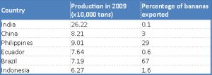 | Table 1- Production and export of banana in 2009. Source- FAOSTAT (2011), Kumar et al. (2010). |