


ISSN : 0976-7061
EISSN : 0976-707X



PARIKH U.R.1*, GOSWAMI H.M.2, NANAVATI M.G.3, BISEN V.V.4, PATEL S.5, MENPARA C.B.6, YADAV K.S.7, SHAH P.K.8, MEHTA N.P.9, GONSAI R.N.10
1Department of Pathology, B.J. Medical College, Ahmedabad-380016, Gujarat, India.
2Department of Pathology, B.J. Medical College, Ahmedabad-380016, Gujarat, India.
3Department of Pathology, B.J. Medical College, Ahmedabad-380016, Gujarat, India.
4Department of Pathology, B.J. Medical College, Ahmedabad-380016, Gujarat, India.
5Department of Pathology, B.J. Medical College, Ahmedabad-380016, Gujarat, India.
6Department of Pathology, B.J. Medical College, Ahmedabad-380016, Gujarat, India.
7Padmashree Dr. D.Y. Patil Medical College and Hospital, Navi Mumbai- 400706, MS, India.
8Professor, Department of Microbiology, B.J. Medical College, Ahmedabad-380016, Gujarat, India.
9Department of Pathology, B.J. Medical College, Ahmedabad-380016, Gujarat, India.
10Department of Pathology, B.J. Medical College, Ahmedabad-380016, Gujarat, India.
* Corresponding Author : urviparikh76@gmail.com
Received : 08-11-2012 Accepted : 29-11-2012 Published : 06-12-2012
Volume : 3 Issue : 2 Pages : 37 - 40
J Clin Res Lett 3.2 (2012):37-40
DOI : http://dx.doi.org/10.9735/0976-7061.3.2.37-40
Conflict of Interest : None declared
Aims and Objective- This study has been undertaken to evaluate the role of Fine Needle Aspiration Cytology (FNAC) in Human Immunodeficiency Virus (HIV) positive lymphadenopathy patients. Materials And Method- Forty HIV positive Patients with lymphadenopathy were subjected to FNAC over a period of eight months i.e. from January, 2011 to August, 2011 in pathology department in our tertiary care hospital. Aspiration was done as a routine procedure using 22 gauge needle with standard precautions after taking detailed clinical history and physical examination of the patients. Smears obtained were stained with May-Grunwald-Giemsa (MGG), Papanicolaou (PAP) and Hematoxylin & Eosin (H&E) stains. Special stain used was Ziehl-Neelsen (ZN) stain for Acid Fast Bacilli (AFB). Result- Age distribution was noted between 5 years to 67 years during the present study. The peak incidence was noted in fourth decade of life (32.5 %). In HIV positive patients, lymphadenopathy showed male predominance, with the male: female ratio of 3.44:1. Cervical lymph nodes were the most common site encountered (62.5 %) as an initial affected site. FNAC results were classified as an inadequate material, non-neoplastic lesions and neoplastic lesions. The most common etiology associated with HIV was Tuberculous lymphadenitis (40.54 %) followed by acute suppurative lymphadenitis (27.03 %). Conclusion- FNAC is simple and safe investigative procedure for diagnosis of lymphadenopathy in HIV positive patients. It obviates surgical excision and guides subsequent therapy and management. Many opportunistic infections can also be found out with the help of this procedure.
Fine Needle Aspiration Cytology (FNAC), Human Immunodeficiency Virus (HIV), Lymphadenopathy.
India has a population of one billion, around half of whom are adults in the sexually active age group. The first AIDS case in India was detected in 1986 and since then HIV infection has been reported in all states and union territories.
The spread of HIV in India has been uneven. Although much of India has a low rate of infection, certain places have been more affected than others. HIV epidemics are more severe in the southern half of the country and the far north-east. The highest estimated adult HIV prevalence is found in Manipur (1.40 %), followed by Andhra Pradesh (0.90 %), Mizoram (0.81 %), Nagaland (0.78 %), Karnataka (0.63 %) and Maharashtra (0.55 %) [1] .
In the southern states, HIV is primarily spread through heterosexual contact. Infections in the north-east are mainly found amongst injecting drug users (IDUs) and sex workers.
Unless otherwise stated, the data on this page has been taken from a 2008 report by the Indian government’s AIDS organization-NACO (National AIDS Control [2] .
Previously it was thought that around 5 million people were living with HIV in India - more than in any other country. Better data, including the results of a national household survey conducted in 2005-2006, led to a major revision of the prevalence estimate in July 2007 [3] . It is now thought that around 2.39 million people in India are living with HIV. Of these, an estimated 39 % are female and 3.5 % are children [1] .
Back-calculation suggests that HIV prevalence in India may have declined slightly in recent years, though the epidemic is still growing in some regions and population groups.
According to NACO, the annual number of AIDS-related deaths peaked in 2006 at 200,000. In 2009 172,000 people were reported to have died from AIDS-related causes [1] .
The National Family Health Survey, which tested more than 100,000 people for HIV, also found prevalence to be higher in urban areas (0.35 %) than in rural areas (0.25 %). It should be noted that many AIDS-related deaths go unreported in India, due to unprecedented levels of stigma and discrimination. In many situations a patient will die without HIV having been diagnosed, and with the death attributed to an opportunistic infection, such as tuberculosis.
Human immunodeficiency virus infection/Acquired immunodeficiency syndrome (HIV/AIDS) is a disease of the human immune system caused by the human immunodeficiency virus (HIV) [4] . During the initial infection a person may experience a brief period of influenza-like illness. This is than typically followed by a prolonged period without symptoms. As the illness progresses it interferes more and more with the immune system, making people much more likely to get infections, including opportunistic infections and tumors that do not usually affect people with working immune systems.
HIV is transmitted primarily via sexual intercourse (including oral sex and anal sex), Contaminated blood transfusions and hypodermic needles and from mother to child during pregnancy, delivery, or breastfeeding [5] . Some bodily fluids, like saliva or tears, do not transmit HIV [6] .Â
HIV is transmitted by three main routes:Â sexual contact, exposure to infected body fluids or tissues and from mother to child during pregnancy, delivery, or breastfeeding which is summarized in given [Table-2] .
Acquired immunodeficiency syndrome (AIDS) is known to be caused by a lymphotropic retro-virus, first described by French investigators and later investigated in United States. AIDS was first recognized in 1981. It has become clear that this syndrome represents the most severe form of a broad spectrum disease [13] . AIDS is a fatal illness that breaks down the body’s immunity and leaves the victim vulnerable to life-threatening opportunistic infections, neurological disorders or unusual malignancies [14] . In India the human immunodeficiency virus (HIV) epidemic is now more than a decade old and has emerged as one of the most serious public health problems in our country [15] . Lymphadenopathy is one of the earliest manifestations of HIV infection, due to the presence and effects of the virus. Lymphadenopathy may also be a manifestation of an opportunistic infections, lymphoid malignancy developing in an immunodeficient individual [16] . Fine needle aspiration cytology (FNAC) can serve as an alternative method and may be practiced for the diagnosis of opportunistic infections in HIV/AIDS patients e.g. tuberculosis, histoplasmosis, toxoplasmosis and malignant conditions such as Kaposi sarcoma and lymphoma [16] . FNAC has become the primary investigative procedure for mass lesions on HIV-positive patients, particularly in the assessment of lymphadenopathy. The procedure is rapid, easily performed and in many cases obviates excision while guiding subsequent therapy or observation. Purpose of the present study is to detect the role of FNAC in diagnosis of lymphadenopathy in HIV positive patients.
A retrospective study of forty cases over a period of eight months i.e. from January, 2011 to August, 2011 was carried out to determine the diagnostic accuracy of FNAC for lymphadenopathy in HIV positive patients, in pathology department of our tertiary care hospital. HIV positivity is confirmed by ELISA test. After detailed clinical history and physical examination, aspiration was carried out using 22 gauge needle and standard precautions. Smears were immediately fixed in 95 % ethyl alcohol. Smears were stained with May Graunwald Geimsa (MGG), Hematoxylin and Eosin stain (H&E) and Papanicolaou (PAP) stain. Special stains used were Zeil -Neilsen (ZN) stain for detecting acid fast bacilli (AFB) and Periodic Acid-Schiff (PAS) for fungi. Cytopathological diagnosis had been recorded in each case.
A total of forty HIV positive patients with peripheral lymphadenopathy diagnosed and treated at our tertiary care hospital over a period of eight months i.e. from January, 2011 to August, 2011 were taken into consideration. In all the cases, pre-operative FNAC was done and diagnosis was recorded. Out of 40 cases, unsatisfactory material was obtained in 3 cases (7.5 %), so no opinion was possible and satisfactory material was obtained in the remaining 37 cases.
Age wise distribution was noted between 5-67 years during the present study. Our youngest patient was 5 year female while the oldest patient was 67 year male. Most of the cases were found in 31-40 years of age (32.5 %) followed by 21-30 years (22.5 %). Out of 40 cases studied, 31 patients were male while 9 patients were female. Peripheral lymphadenopathy showed male predominance- with the male to female ratio 3.44:1.
The most common site of peripheral lymphadenopathy was the cervical group; consist of 25 out of 40 cases (62.5 %).
During cytological examination; tuberculous lymphadenopathy (40.54 %) was the most common lesion encountered during the present study (15 out of 37 patients). Out of them, 6 patients had caseous necrosis and granuloma, 4 patients had granuloma without caseous necrosis and 5 patients had caseous necrosis without granuloma formation. All the patients underwent AFB stain. Seven patients (46.67 %) showed positive AFB staining.
A total of 10 cases (27.03 %) showed acute suppurative lymphadenitis as they showed plenty of acute inflammatory infiltrate on necrotic background. ZN stain and PAS stain were negative.
One patient showed neoplastic proliferation of small lymphocytes with minimal pleomorphism and it was reported as Non-Hodgkin’s Lymphoma (2.7 %). Smear was suggestive of diffuse large B cell lymphoma though further typing was not possible. No opportunistic infection other than mycobacterium tuberculosis was found in the patients examined during the present study.
Seven patients (18.92 %) showed reactive lymphadenitis while four patients (10.81 %) showed cytomorphology of chronic non-specific lymphadenitis. All of them were negative for AFB screening by ZN staining.
HIV is more prevalent in developed countries than India. Even in India, as the incidence of HIV is increasing day by day, complications are also rising continuously. And so it is important to consider the detail study of lesions associated with HIV infection. Peripheral lymphadenopathy is one of the common conditions associated with HIV infection. FNAC is regarded as the gold standard initial investigation in the diagnosis of condition responsible for peripheral lymphadenopathy. FNAC is safe, simple, cost effective screening test for rapid diagnosis and have low complication rate [16] .
During present study, age distribution was noted between 5 years to 67 years. The commonest age group to be affected was fourth decade of life (32.5 %) followed by third decade (22.5 %). During his study Bates, et al [17] . found 22 males and 1 female HIV positive patients were presented with lymphadenopathy and their age ranged from 19 to 72 years. Further, cervical lymph nodes were the most commonly affected site. During the present study we found males are predominantly affected by HIV infection with the male: female ratio of 3.44:1. Shenoy, et al [18] . found the male: female ratio was 5:1 and the maximum numbers of the patients were present in 25-30 years of age group. Cervical group of lymph nodes being the most commonly affected site.
In the present study as well as study performed by Shenoy, et al [18] . Mycobacterial infection was found to be more common among all the infectious diseases; because of increased prevalence of mycobacterial tuberculosis in our country than in developed country. Over and above this; decreased immunity due to HIV infection is also responsible for the same.
Out of all the tuberculous patients, we found 40 % (6 patients) had caseous necrosis with granuloma, 26.67 % (4 patients) had granuloma without caseous necrosis and 33.33 % (5 patients) had caseous necrosis only. In the study conducted by Vanisri, et al [19] . Caseous necrosis with epitheloid granuloma was observed in 50 % of cases. Granuloma without caseous necrosis was observed in 30 % of cases and only caseous necrosis was observed in 20 % of cases. Thus the results are comparable. In the study performed by Vanisri, et al [19] . one case of Mycobacterium avium intercellulare (MAI) lymphadenitis was reported that showed aggregates of pale histiocytes with foamy cytoplasm in the smears with poorly formed granuloma. The smear stained positive for both AFB and PAS, though MAI was not found in our study group.
In the present study, ten cases (27.03 %) were found to have acute suppurative lymphadenitis. ZN and PAS stain were negative in these cases. Smear showed acute inflammatory infiltrate on necrotic background. In the study performed by Shenoy, et al [18] . acute Suppurative lymphadenitis with AFB positivity was observed in 3 (13 %) patients. In the study performed by Bates, et al and Reid, et al showed 7.4 % and 7 % patients had acute suppurative infection respectively [17,20] . The higher number of abscess during the present study compare to Bates, et al and Reid, et al is due to decreased immunity and increased risk of infection in HIV positive patients in our country.
Seven cases (18.92 %) showed reactive lymphadenitis in present study. Smears showed polymorphous cell population with mature and transformed lymphocytes, monocytoid cells, neutrophils and tingible body macrophages. In the study conducted by shenoy, et al [18] . 35.7 % of patients showed reactive lymphadenitis. Bates, et al found reactive hyperplasia in 41 % of aspirates [17] and Reid, et al found reactive hyperplasia in 51 % of aspirates [20] . Our results were quite lower compare to Bates, et al and Reid, et al is due to immune-compromised status in HIV positive patients. All cases of reactive lymphadenitis in present study were negative for AFB and PAS stain. The difference is due to early diagnosis and availability of antiretroviral therapy in HIV positive patients.
In the present study, chronic non-specific lymphadenitis was found in 4 cases (10.81 %). In study conducted by Shenoy, et al. 4.5 % cases were diagnosed as chronic non-specific lymphadenitis [18] . During their study Bates, et al and Reid, et al showed 10.6 % and 20 % cases were diagnosed as chronic non-specific lymphadenitis respectively [17,20] . Our result is comparable with Bates, et al and Reid, et al.
Non-hodgkin’s lymphoma is one of the commonest malignancies encountered in HIV associated lymphadenopathy. One case (2.7 %) showed features of NHL, with smear showing possibility suggestive of diffuse large B cell lymphoma. In study by Shenoy, et [18] 8.9 % showed lymphoma, with Bates, et al [17] and Reid, et al [20] showing 4 % and 5 % respectively. Kaposi sarcomas were found in western literature, [17,20] though it was not found in our study.
In the present study, evidence of opportunistic infections other than Mycobacterium tuberculosis was not found in any of the lymph nodes examined. In the study performed by Bates, et al [1] case of Histoplasma and one case of Cryptococcus were found.
FNAC is the primary and safe investigative procedure for lesions of lymph nodes in HIV patients. Procedure is rapid and easily performed it obviates the need of excision, guides subsequent therapy or observation and provide definite guidelines for management. It is one of the sensitive as well as specific diagnostic tests with minimum complications.
[1] NACO (2011) Annual Report -2010-2011.
» CrossRef » Google Scholar » PubMed » DOAJ » CAS » Scopus
[2] NACO (2008)Â HIV Sentinel Surveillance and HIV Estimation in India 2007: A Technical Brief.
» CrossRef » Google Scholar » PubMed » DOAJ » CAS » Scopus
[3] WHO (2007)Â 2.5 Million People in India Living with HIV, According to New Estimates.
» CrossRef » Google Scholar » PubMed » DOAJ » CAS » Scopus
[4] Sepkowitz KA (June 2001) N. Engl. J. Med., 344(23), 1764-72.
» CrossRef » Google Scholar » PubMed » DOAJ » CAS » Scopus
[5] Markowitz (2007) Environmental and Occupational Medicine (4th ed.).
» CrossRef » Google Scholar » PubMed » DOAJ » CAS » Scopus
[6] Centers for Disease Control and Prevention (2003) HIV and Its Transmission.
» CrossRef » Google Scholar » PubMed » DOAJ » CAS » Scopus
[7] Smith D.K. Grohskopf L.A. Black R.J. Auerbach J.D. Veronese F., Struble K.A., Cheever L., Johnson M., Paxton L.A., Onorato I.M., Greenberg A.E. (2005) U.S. Department of Health and Human, Services.
» CrossRef » Google Scholar » PubMed » DOAJ » CAS » Scopus
[8] Coovadia H. (2004) N. Engl. J. Med., 351(3), 289-292.
» CrossRef » Google Scholar » PubMed » DOAJ » CAS » Scopus
[9] Kripke C. (2007)Â American Family Physician, 76(3), 375-6.
» CrossRef » Google Scholar » PubMed » DOAJ » CAS » Scopus
[10] Dosekun O., Fox J. (2010) Current Opinion in HIV and AIDS., 5(4) 291-7.
» CrossRef » Google Scholar » PubMed » DOAJ » CAS » Scopus
[11] Boily M.C., Baggaley R.F., Wang L., Masse B., White R.G., Hayes R.J., Alary M. (2009) The Lancet Infectious Diseases 9(2), 118-29.
» CrossRef » Google Scholar » PubMed » DOAJ » CAS » Scopus
[12] Baggaley R.F., White R.G., Boily M.C. (2008) International Journal of Epidemiology, 37(6), 1255-65.
» CrossRef » Google Scholar » PubMed » DOAJ » CAS » Scopus
[13] Ewing E.P., Chandler F.W., Spira T.J., Brynes R.K., Chan W.C. (1985) Arch. Pathol. Lab. Med., 109, 977-81.
» CrossRef » Google Scholar » PubMed » DOAJ » CAS » Scopus
[14] Prasad H.K., Bhojwani K.M., Shenoy V., Prasad S.C. (2006) Am. J. Otolaryngol, 27, 179-85.
» CrossRef » Google Scholar » PubMed » DOAJ » CAS » Scopus
[15] Joshi P.L., Rao J.V. (1999) AIDSÂ Res. Rev., 2, 7-9.
» CrossRef » Google Scholar » PubMed » DOAJ » CAS » Scopus
[16] Shobhana A., Guha S.K., Mitra K., Dasgupta A., Negi D.K., Hazra S.C. (2002) Indian J. Med. Microbiol., 2, 99-101.
» CrossRef » Google Scholar » PubMed » DOAJ » CAS » Scopus
[17] Martin-Bates E., Tanner A., Suvarna S.K., Glazer G., Coleman D.V. (1993) J. Clin. Pathol., 46, 564-6.
» CrossRef » Google Scholar » PubMed » DOAJ » CAS » Scopus
[18] Shenoy R., Kapadi S.N., Pai K.P., Kini H., Mallya S., Khadilkar U.N., et al. (2002) Acta. Cytol., 46, 35-9.
» CrossRef » Google Scholar » PubMed » DOAJ » CAS » Scopus
[19] Vanisri H.R., Nandini N.M., Sunila R. (2008) Indian J. Pathology & Microbiology, 51(4), 481-484.
» CrossRef » Google Scholar » PubMed » DOAJ » CAS » Scopus
[20] Reid A.J., Miller R.F., Kocjan G.L. (1998) Cytopathology, 9, 230-9.
» CrossRef » Google Scholar » PubMed » DOAJ » CAS » Scopus
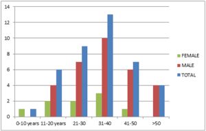 | Fig. 1- Age and sex wise distribution of lymphadenopathy in HIV positive patients |
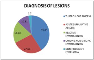 | Fig. 2- Diagnosis of lymphadenopathy in HIV positive patients |
 | Table 1- HIV data from National AIDS Control Organization |
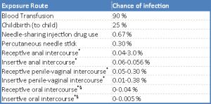 | Table 2- Average per act risk data of getting HIV by exposure route to an infected source *assuming no condom use, § source refers to oral intercourse performed on a man |
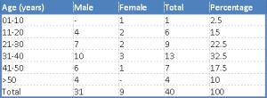 | Table 3- Age and Sex Wise Distribution of Lymphadenopathy in HIV Positive Patients |
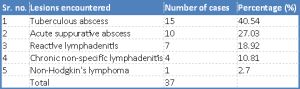 | Table 4- Diagnosis of lymphadenopathy in HIV positive patients |
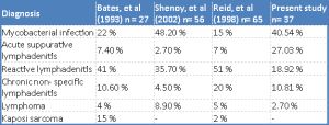 | Table 5- Comparison of results of FNAC studies in HIV positive patients |