ISSN : 0976-5530
EISSN : 0976-5549

GANDHAM N.R.1*, SINGH G.2, ROY I.3, VYAWAHARE C.4, GOOPTU S.5, JADHAV S.V.6, MISRA R.N.7
1Department of Microbiology, Pad. Dr. D.Y. Patil Medical College, Hospital and Research Centre, Pune- 411018, MS, India.
2Department of Surgery, Pad. Dr. D.Y. Patil Medical College, Hospital and Research Centre, Pune- 411018, MS, India.
3Department of Microbiology, Pad. Dr. D.Y. Patil Medical College, Hospital and Research Centre, Pune- 411018, MS, India.
4Department of Microbiology, Pad. Dr. D.Y. Patil Medical College, Hospital and Research Centre, Pune- 411018, MS, India.
5Department of Surgery, Pad. Dr. D.Y. Patil Medical College, Hospital and Research Centre, Pune- 411018, MS, India.
6Department of Microbiology, Pad. Dr. D.Y. Patil Medical College, Hospital and Research Centre, Pune- 411018, MS, India.
7Department of Microbiology, Pad. Dr. D.Y. Patil Medical College, Hospital and Research Centre, Pune- 411018, MS, India.
* Corresponding Author : nageswari_rg@rediffmail.com
Received : 22-01-2013 Accepted : 31-01-2013 Published : 01-07-2013
Volume : 4 Issue : 1 Pages : 242 - 244
Int J Med Clin Res 4.1 (2013):242-244
DOI : http://dx.doi.org/10.9735/0976-5530.4.1.242-244
Conflict of Interest : None declared
Corynebacteria other than C. diphtheriae are increasingly being implicated as pathogens. C. ulcerans, C. urealyticum, C. amycola-tum, C. striatum are some of the species being increasingly identified and implicated as pathogens. C. striatum has been associated with wound infections, respirarory tract infections and foreign body infections. Further it can establish itself as a nosocomial pathogen. We describe a case of Corynebacterium striatum, necrotizing fasciitis in a HBsAg positive patient, with history of trauma. The bacterium was considered etiologically significant as the gram smear from the sample showed numerous gram positive bacilli & culture rendered pure growth of C. stria-tum. The isolate was identified by the Vitek 2C automated ID system from Biomeriux. Patient was treated by the culture-sensitivity report. Fol-lowing which he needed a skin graft. He was discharged with complete recovery. C. striatum is often implicated as pathogen in immunocom-promised and now also in immunocompetent patients.
Coryneforms, C. striatum, identification, Necrotising fasciitis
Coryneform bacteria includes as a group of bacteria belonging to different genera mainly sharing characteristics like being Gram positive rods with club- shaped ends, V-shaped with one end in contact, pallisading pattern in morphology, non- motile and in the biochemicals being catalase positive and oxidase negative. In this group the genus Corynebacterium is the most important medically with C. diphtheriae being the single most prominent pathogen. Most of the Corynebacterium other than C. diptheriae were referred to as “diptheroids†often considered as a part of normal skin commensal flora of human being [1] . Several Corynebacterium species C. jeikeium, C. urealyticum, C. ulcerans have gained medical interest in community acquired and nosocomial infections due to improved isolation-identification leading to an evolving number of publications and better clinical correlation [2] . Corynebacteria have been implicated as opportunistic pathogen in setting of different immunocompromised conditions like diabetes & viral infections, eg. Herpes genitalis [3] . Being part of normal skin flora C. striatum is associated with wound infections, respiratory tract infections and foreign body infections. Herein we report a case of C. striatum infection and discuss its diagnosis, treatment and review its role as a pathogen.
A 37 year old male patient presented in Surgery OPD of Pad. Dr. D.Y. Patil Medical College, Hospital, Pune with diffuse swelling of the left lower limb of five days duration which developed following blunt trauma. He was non diabetic but was detected to be HBsAg positive. There were no constitutional symptoms. He was diagnosed as a case of cellulitis and started on combination of crystalline penicillin, Gentamicin and Metrogyl alongwith other supportive measures. Over next four days swelling increased and overlying skin showed blackish discoloration. Diagnosis was revised to Necrotizing fasciitis and debridement was done.
A sample taken from the lesion was sent for microbiological investigation. Culture, grew Corynebacterium species & Pseudomonas aeruginosa. Haematological findings revealed polymorphonuclear leucocytosis (TLC of 12,000). The patient was treated with multiple courses of antibiotics including carboxypenicillin, third generation cephalosporin, gentamycin. Wound perfusion was delayed over two months because of infection. The patient was subsequently reviewed for microbiologic examination, at which time careful re-sampling of the lesion was done.
On examination the wound occupying 3/4th entire left leg below knee and above ankle was seen [Fig-1] . Gram stained smears of discharge of the ulcerative lesion showed overwhelming presence of Gram positive bacilli in palisade formation. Many of the bacilli were visualized within and outside polymorphonuclear leukocytes [Fig-2] . Inoculation of the sample (pus) onto Blood Agar grew pure cultures of white to cream coloured, non-haemolytic, smooth colonies [Fig-3] . Gram stained smears of growth showed Gram positive bacilli V-shaped, some of which upon careful evaluation showed a stripped pattern with clubbed ends [Fig-4] . They were and non-motile, positive for catalase test, nitrate reduction test, glucose, sucrose fermentation test and negative for urease test [4] . Isolate was confirmed as C. striatum through Vitek-2C automated system from Biomeriux, using ANC card. The salient reactions were glucose, sucrose fermentation, nitrate reduction and negative urease production. Antimicrobial susceptibility was performed by Kirby-Bauer Disk Diffusion method. The isolate was susceptible to vancomycin, imipenem, linezolid, penicillin, gentamycin and resistant to cotrimoxazole, erythromycin, clindamicin, tetracycline [5,6] . Haemotological findings at this time revealed marked polymorphonuclear leucocytosis (TLC 25000), Hb 13.5gm%, DLC P84, L12, E02, M02. Gram stained smears & culture were repeated and were positive for C. striatum.
Antibiotics as per culture and sensitivity were administered. Resultant healthy wound occupying almost 3/4th circumference of entire leg sparing ankle was covered with split skin graft. Graft take was almost 90% and the wound healed completely thereafter.
For many years C. striatum was long believed to have limited potential as a pathogen and hence was usually considered as a contaminant when isolated from a patient sample. In 1980, the first case of human pleuro-pulmonary infection with C. striatum was reported in a 79 year old man with chronic lymphocytic leukaemia [7] . In a series reported by Martinez-Martinez et.al. 26 patients with positive C. striatum cultures were identified as having significant infections as defined by CDC criteria. In seven of 26 patients, the organism was isolated from a culture of a chronic skin ulcer and another seven had surgical wound infections which provide evidence of C. striatum as one of the causative organisms of chronic cutaneous ulcer [8] .
Evidence to support the role of C. striatum as a pathogen in immunocompromised as well as immunocompetent hosts is growing. The role of C. striatum as a nosocomial pathogen is also evolving as it has been reported in several hospital outbreaks [9] . As a colonizer of human skin C. striatum can establish de novo cutaneous infections through disruption of intact skin barriers as in our case, or can invade preexisting cutaneous lesion. Watkins et.al. in his series of six cases include one of 78 year old woman with history of colorectal carcinoma, who punctured her finger on a rose thorn and developed a pyogenic granuloma. Culture of the biopsy material revealed a heavy growth of C. striatum and Ps. aeruginosa [10] . Peiris et.al. described a 73 year old women with peripheral vascular disease who had a deep skin sinus around her elbow, which was oozing pus. The sinus tract had been present for months and developed in absence of overt trauma. Culture of the exudates showed heavy growth of C. striatum [11] . A similar case of C. striatum infected ischaemic ulcer in a72 year old diabetic was reported by Martinez-Martinez [8] . Martin et.al. reported a case of C. striatum in a 69 year old patient with chronic ischaemia of the lower extremites and lesions in the first, second toe and heel of the right foot [12] . In our patient, it is conceivable that his deep seated infection was attributable to an incident induced by a trauma, which could have provided the portal of entry for C. striatum perhaps colonizing his skin. Furthermore, Superti et.al. reported a case of C. striatum skin and soft tissue infection of a malignant skin lesion in a 27 year old male patient indicating that this bacterium has a predilection for devitalised cutaneous and soft tissue [13] . Most C. striatum infections take place in patients with underlying medical conditions.
C. striatum infections involving the skin have also been reported in normal hosts [14] . Stone et.al reported a case of recurring breast abscess that required several drainage procedures over a seven week period in a 41 year old immune competent woman with no underlying medical condition [15] . Microscopy of the devitalized tissue showed large number of Gram positive bacilli and pure growth on culture. From the different published cases it seems that most but not all patients have been hospitalized and have had an underlying or associate diseases. In this report, the majority of patients had been hospitalized for many days as studied by Rizvi M et.al. [16] .
Corynebacterium should be identified to species level if they are isolated from- 1) normally sterile sites. 2) from adequately collected clinical material if they are predominant organisms and total bacterial count is >10 5/ml. Further positive multiple specimen for same bacteria and positive direct microscopy along with strong leukocyte reaction strengthens it’s clinical significance [4] .
These criteria helped establish the role of C. striatum as a pathogen in this patient. It is conceivable that his fasciitis was attributable to an incident induced by a trauma, leading to devitalized tissue which provided the portal of entry for C. striatum perhaps colonizing his skin.
Isolation of C. striatum from a clinical specimen in a patient with suspected infection should not be ignored. Evidence of large number of organisms on Gram stain or predominant growth in culture or bacteremia support its role as a potential pathogen. The antibiotic susceptibility pattern of C. striatum is variable and empiric therapy with a glycopeptides should be considered while awaiting the susceptibility pattern of a particular isolate. The role of C. striatum as a nosocomial pathogen needs to be kept in mind while identifying and reporting this isolate from patient samples. The reason being that often the first isolates is followed by a cluster of isolates from the same ward or surgical unit. Hence an alert microbiologist can play a major role in arrest and control of such a cluster of cases.
C. striatum is now an established pathogen, both in immunocompromised and immunocompetent patients. It is increasingly being reported to cause infection in long standing open wounds. Isolation of C. striatum from a clinical specimen in a patient with suspected infection should not be ignored in view of its propensity to establish nosocomial infections. Evidence of large number of organisms on Gram stain or predominant growth in culture or bacteremia, support its role as a potential pathogen. Antibiotic therapy with a glycopeptide while awaiting culture- sensitivity report of a particular isolate could benefit the outcome. This along with appropriate measures can limit its role as a nosocomial pathogen.
[1] Bottone E.J., Fabbri M., Ashraf A. (2010) Reviews in Infection, 1(2), 104-109.
» CrossRef » Google Scholar » PubMed » DOAJ » CAS » Scopus
[2] Martinez-martinez L., Suarez AI, Winstanley J., Ortega M.C., Bernard K. (1995) J. Clin. Microbiol., 33, 2458-2461.
» CrossRef » Google Scholar » PubMed » DOAJ » CAS » Scopus
[3] Ghosh P., Mangal K.K., Sharma Y.K., Misra R.N., Dash K.D. (2012) Journal of Clinical and Diagnostic Research, 6(7), 1298-1300.
» CrossRef » Google Scholar » PubMed » DOAJ » CAS » Scopus
[4] Funke G., Barnard K.A. (2007) Manual of Clinical Microbiology. 9th ed., Washington: ASM Press, 485-514.
» CrossRef » Google Scholar » PubMed » DOAJ » CAS » Scopus
[5] Martinez-martinez L., Pascual A., Bernard K., Suarez A.I. (1996) Antimicrob. Ag. Chemother., 40, 2671-2672.
» CrossRef » Google Scholar » PubMed » DOAJ » CAS » Scopus
[6] Reddy B.S., Chaudhury A., Kalawat U, Jayaprada R., Reddy G.S.K., Ramana B.V. (2012) Indian Journal of Medical Microbiology, 30(1), 52-57.
» CrossRef » Google Scholar » PubMed » DOAJ » CAS » Scopus
[7] Bowsted I.I., Santiago S.M. Jr. (1980) Br. J. Dis. Chest, 180, 74, 198-200.
» CrossRef » Google Scholar » PubMed » DOAJ » CAS » Scopus
[8] Martinez-martinez L., Suarez A.I., Rodriguez-Bano J., Bernard K., Muniain M.A. (1997) Clinic. Micro. & Inf., 3(6), 634-39.
» CrossRef » Google Scholar » PubMed » DOAJ » CAS » Scopus
[9] Funke G., Graevenitz A.V., Clarridge J.E. & Bernard K.A.(1997 Clin. Microbiol. Rev., 10, 125-159.
» CrossRef » Google Scholar » PubMed » DOAJ » CAS » Scopus
[10] Watkins D.A., Chahine A., Richard C.J., Jacobs M.R., Lazarus H.M. (1993) Clinical Infectious Diseases, 17, 21-25.
» CrossRef » Google Scholar » PubMed » DOAJ » CAS » Scopus
[11] Peiris V., Fraser S., Knowles C., Norris S., Bennet C. (1994) Eur. J. Clin. Microbiol. Infect. Dis., 13, 36-8.
» CrossRef » Google Scholar » PubMed » DOAJ » CAS » Scopus
[12] Martin M.C., Melon O., Celada M.M., Alvarez J., Mendez F.Z., Vazquez F. (2003) Journal of Medical Microbiology, 52(7), 599-602.
» CrossRef » Google Scholar » PubMed » DOAJ » CAS » Scopus
[13] Superti S.V., Martins D.S., Caierao J., Soares F., Procnow T., Cantarelli V.V., Zavascki A.P. (2009) Inst. Med. Trop. S. Paulo, 51(2).
» CrossRef » Google Scholar » PubMed » DOAJ » CAS » Scopus
[14] Lee P.P., Ferguson Jr. D.A., Sarubbi F.A. (2005) J. Infect., 50, 338-343.
» CrossRef » Google Scholar » PubMed » DOAJ » CAS » Scopus
[15] Stone N., Gillett P., Burge S. (1997) British Journal of Dermatology, 137(4), 623-625.
» CrossRef » Google Scholar » PubMed » DOAJ » CAS » Scopus
[16] Rizvi M., Khan F., Raza A., Shukla I., Malik A., Rizvi S.A.R., Sherwani M.K., Afzal K. and Hasan S.A. (2011) American-Eurasian Journal of Scientific Research, 6(3), 165-171.
» CrossRef » Google Scholar » PubMed » DOAJ » CAS » Scopus
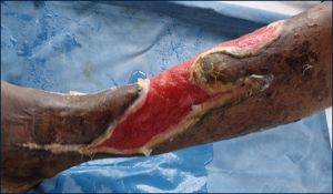 | Fig. 1- spreading lesion on leg |
 | Fig. 2- Gram stain of discharge |
 | Fig. 3- Colonies on blood agar |
 | Fig. 4- Gram stain of growth |