ISSN : 0976-5530
EISSN : 0976-5549

MATNANI G.1, ROY I.2, GANDHAM N.3, UJAGARE M.4, JADHAV S.V.5*
1Department of Microbiology, Pad. Dr. D.Y. Patil Medical College and Hospital, Pimpri, Pune-411018, MS, India.
2Department of Microbiology, Pad. Dr. D.Y. Patil Medical College and Hospital, Pimpri, Pune-411018, MS, India.
3Department of Microbiology, Pad. Dr. D.Y. Patil Medical College and Hospital, Pimpri, Pune-411018, MS, India.
4Department of Microbiology, Pad. Dr. D.Y. Patil Medical College and Hospital, Pimpri, Pune-411018, MS, India.
5Department of Microbiology, Pad. Dr. D.Y. Patil Medical College and Hospital, Pimpri, Pune-411018, MS, India.
* Corresponding Author : patilsv78@gmail.com
Received : 17-09-2012 Accepted : 03-10-2012 Published : 29-10-2012
Volume : 3 Issue : 7 Pages : 215 - 220
Int J Med Clin Res 3.7 (2012):215-220
DOI : http://dx.doi.org/10.9735/0976-5530.3.7.215-220
Conflict of Interest : None declared
Introduction- Fungi are world- wide in distribution but only few of them are considered as pathogenic. As the fungal infections are not notifiable infections like viral, parasitic, bacterial diseases hence these are not given much attention and usually the diagnosis is established very late. In response to the increased incidence of fungal infections, the pharmaceutical industry has developed a number of newer less toxic anti-fungal drugs for clinical use. The increased use of antifungal drugs, often for prolonged periods, has led to acquired antifungal resistance among previously susceptible strains or species and to the increased incidents of infections with less common species. Objective- To study the fungal isolates in relation to site of infection/specimen and to establish in vitro anti-fungal susceptibility testing. Materials and Methods- The study was conducted during January 2007 to December 2008 Pad. Dr. D.Y. Patil Medical College and Hospital Pimpri, Pune-18. Total 120 clinically suspected cases of superficial fungal infections were collected for study from various wards and OPDs of hospital. Results and Observation- Superficial fungal infection occurs mainly in younger age group and adults. In the present study males were more affected than females. Most common clinically diagnosed cases in superficial fungal infection of skin were of Tinea corporis, T. pedis and T. cruris. Among dermatophytes, T. rubrum was the commonest etiological agent followed by T. mentagrophytes. Amongst the non-dermatophyte moulds, Aspergillus spp. was the most prevalent species. SAAS method showed potential as a screen for anti-fungal susceptibility testing. Anti-fungal susceptibility testing of dermatophytes (Trichophyton rubrum was done by semi-solid agar dilution method using ant-fungal drug terbinafine. All isolates were sensitive. MIC of all isolates were 0.25µg/ml. Conclusion- To reduce the time required for anti-fungal susceptibility testing of filamentous fungi, semi-solid agar susceptibility method uses inoculums suspensions that can be readily prepared from the original pure plate. The test can even set up as soon as the mould is isolated. No special expertise or expensive equipments is needed, because the procedure is simple and similar for all fungi.
Superficial fungal infections, Antifungal susceptibility, Trichophyton rubrum, Candida albicans.
Fungi are world- wide in distribution but only few of them are considered as pathogenic. As the fungal infections are not notifiable infections like viral, parasitic, bacterial diseases hence these are not given much attention and usually the diagnosis is established very late. The approach to fungi identification in the developing countries is on the gross morphological features whereas in the developed countries it is the molecular approach. The incidence of fungal infections has increased dramatically in the past 20 years partly because of the increase in the number of people whose immune systems are compromised by either aging or as acquired immunodeficiency syndrome (AIDS), organ transplantation or cancer therapy [1-3] . Accordingly, the increase in rates of morbidity and mortality, due to fungal infections has been now recognized as a major problem. However, the epidemiological features of these diseases are not well documented. The pathogenic fungi may give rise to infections in animals and human beings. Most of the agents cause infection of the superficial layers of the integument and only very few give rise to systemic involvement. The superficial cutaneous infections involve the outer most layer of the skin and its appendages like hair and nails [4-6] . These are amongst the most commonly encountered infectious diseases prevalent in most parts of the world. The causative agents colonize cornified layers of epidermis or supra-follicular portions of the hair and do not penetrate into deeper anatomical sites. The patients invariably neglect such type of infections and seek medical attention for cosmetic reasons. Oral anti-fungal drugs, as effective as topical therapy, are preferred by most of the clinicians [1-6] .
In response to the increased incidence of fungal infections, the pharmaceutical industry has developed a number of newer less toxic anti-fungal drugs for clinical use. The increased use of antifungal drugs, often for prolonged periods, has led to acquired antifungal resistance among previously susceptible strains or species and to the increased incidents of infections with less common species. Concurrent with the increase in fungal infections, a large variety of anti-fungal drugs are available with different spectrum of activity. There is therefore a need to determine the anti-fungal susceptibility of isolates to available drugs. Newer anti-fungal agents have increased the therapeutic options thereby leading to the demand for in vitro determination of anti-fungal susceptibility [6-9] . But the application of in vitro anti-fungal susceptibility testing in clinical research and to guidance to anti-fungal therapy has been limited by a lack of reproducibility and uncertain clinical relevance. Unlike anti-bacterial susceptibility testing, anti-fungal susceptibility testing is still in its infancy [8-10] . Although considerable work remains to be done, routine susceptibility testing of fungi will become meaningful for clinical decision making in the foreseeable future. Hence present was taken to isolate and identify the fungal strains from suspected clinical samples and to study the fungal isolates in relation to site of infection and to establish in vitro anti-fungal susceptibility testing.
Study Area- The study was conducted during January 2007 to December 2008 at Pad. Dr. D.Y. Patil Medical College and Hospital Pimpri-Pune-18 India.
Ethics Statement- Written informed consents were obtained from all patients and study protocol were approved by the institutional ethical committee of Dr. D.Y. Patil Medical College, Pune.
Sample Size- Total 120 clinically suspected cases of superficial fungal infections were collected for study from various wards and OPDs of the hospital.
Scraping from Skin- The part to be scraped was cleaned with the swab of 70% alcohol. Sterile scalpel blade was used for scraping. The scraping was done from active peripheral margin of the lesion without injuring the skin surface [11-20] .
Scraping from Nail- The affected nail becames opaque, brittle, hypertrophied. Site of the nail was cleaned with 70% alcohol. Deeper fragments which included crusty deposits from the junction of nail were collected with the help of sterile scissors or nail clippers. These were used for microscopy as well as culture [11-20] .
All culture slants were examined routinely for presence of fungal growth with respect to the colonial appearance like size, surface, color, margin, texture, diffusion of pigmenting the media. Findings were recorded weekly for a period of 3 weeks & negative cultures were discarded after 3 weeks [11-20] .
Microscopic morphology of fungi positive culture slants were studied by following methods [12-15] .
• By Wet Mount- a small portion of the colony of the test fungi was placed in a drop of saline. This preparation was teased & observed under microscope for morphological study of fungi [11-15] .
• By Lactophenol Cotton Blue- for this method, lactophenol cotton blue stain was used. A small portion of a colony was picked and suspended in a solution of lactophenol cotton blue on a slide. The mycelia mat was teased apart with dissecting needles, covered with cover-slip and observed under microscope for presence of hyphae, conidia & manner in which the conidia were attached to the hyphae [11-15] .
• Slide Culture Technique- was used to study morphological details of fungi particularly, relationship between reproductive structures like conidia, conidiophores, hyphae [11-15] .
• Dermatophyte Test Media (DTM)- This medium was used to confirm whether the fungus grown was dermatophyte. This medium indicated growth of dermatophytes with color changes of the medium from yellow to red. All species of dermatophytes showed this color change. The slopes were inoculated with the fungal growth & incubated after isolation of dermatophytes [12-17] .
• Hair Perforation Test- This test was done to differentiate between T. mentagrophytes and T. rubrum. A filter paper strip was placed into bottom of a sterile petridish & cover surface of strip was covered with distilled water. A small portion of sterilized hair was added into the water. Colony was inoculated directly on hair & incubated at 25°C for 10 to 14 days. Hair was observed regularly by making a wet smear for presence of conical perforation of hair-shaft. In contrast to T. rubrum, T. mentagrophytes perforated the hair [12-17] .
• Urease Test- Modified Christensen’s urea Agar-was inoculated with fungal growth. Trichophyton mentagrophytes produced urease within 2 to 3 days after inoculation. T. mentagrophytes hydrolysed urea & changed the color from yellow to pink [12-17] .
1. Germ Tube Test- 0.5 ml of human serum was inoculated with isolated yeast colony. It was incubated for 2 hours at 35°C. After incubation one loopful of yeast inoculated serum was taken on dry, clean slide & covered by cover-slip and observed under low and high magnification for germ tubes. If the test was positive, presumptive identification of C. albicans was made [10-14] .
2. Chlamydospore Formation- An isolated colony from the primary culture medium was obtained. The plate of cornmeal agar was inoculated by making three parallel streaks about ½-inch apart at a 45о angle to the culture medium. Formation of large, highly retractile, thick walled, terminal spore was called chlamydospore. The test was used for the identification of C. albicans [10-14] .
Method-Mueller- Hinton agar supplemented with 2% glucose and 0.5μg methylene blue/ml. Depth 4mm was used. Inoculum-the agar surface was inoculated by using a swab dipped in a cell suspension adjusted to the turbidity of 3.0 McFarland standards. C. albicans ATCC90029 used as control strain. Anti-fungal disc-fluconazole (25μg) disc placed onto the surface of the inoculated plates. Incubation-plates were incubated at 37°C for 24 hrs.
Interpretive Criteria- Susceptible(S)-Zone diameter≥19mm. Susceptible dose dependent (SDD)-Zone diameter-(15-18) mm. Resistance (R)- Zone diameter≤14mm [12-17] .
Semisolid Agar Anti-fungal Susceptibility (SAAS) Testing of Isolated Dermatophytes- Trichophyton rubrum. Media -Sabouraud’s Dextrose broth with 0.5% agar. Drug- terbinafine (0.03-1) μg/ml- stock solution prepared in DMSO (dimethyl sulfoxide). Inoculums-Prepared for filamentous fungi (Trchophyton rubrum) grown on Potato dextrose agar. Swabbing the pure colony (mixture of conidia & hyphal fragments) was suspended in (3-4)ml. of sterile saline. Mixture was vortexed & heavy particles were allowed to settle. Homogenous suspension adjusted to 3.0 McFarland turbidity standard (T. rubrum ATCC1546 used as control strain). Method-Semi solid agar tubes containing known concentrations of terbinafine drug as well as drug free control. Inoculated one loopful of 3.0 McFarland adjusted culture by inserting the loop deep within the semisolid agar. Incubation-tube incubated at 37°C for 96 hrs. End point determination-According to CLSI guideline- Growth compared with drug free control scored by visual inspection [21-25] .
4+ growth same as control
3+ slight decrease in growth
2+ significant reductions in growth
1+ slight growth or few visible hyphal fragments
0- No growth
Semisolid agar anti-fungal susceptibility testing (SAAS) of isolated Aspegillus fumigatus and Aspergillus niger) Media-Sabouraud’s Dextrose broth with 0.5% agar. Drug- Itraconazole- stock solution prepared in DMSO (Dimethyl Sulfoxide) Inoculums [21-25] .
Prepared for filamentous fungi (Aspergillus spp.) grown on potato dextrose agar. Swabbing the pure colony (mixture of conidia & hyphal fragments) was suspended in (3-4) ml. of sterile saline. Mixture was vortexed & heavy particles were allowed to settle. Homogenous suspension adjusted to 3.0McFarland turbidity standard (Aspergillus fumigatus ATCC 204304 and A. niger ATCC16404 used as control strain) [21-25] .
A total of 120 clinically suspected cases of superficial fungal infections were studied. Out of 120 cases, 44 cases were superficial fungal infection of skin. Age and sex wise distribution, of different clinical types of superficial fungal infections of skin, shown in [Table-1] and [Table-2] :
Most common cases were Tinea corporis, followed by Tinea pedis and Tinea cruris. Most common affected age group-21-30 years and above 50 years [Table-1] . Males were more affected than females [Table-2] . Out of 44 cases, 30 cases are KOH positive and 14 cases were KOH negative [Table-3] . Out of 44 cases, 8 cases were culture positive [Table-4] .
All culture positive isolates were Trychophyton rubrum. 18.18% cases were both KOH and Culture positive,50% cases are KOH positive but culture negative, there was no case of KOH negative and culture positive,31.81% cases were both KOH and Culture negative [Table-5] . Total 6 cases of Tinea capitis were found. Out of 6 cases,3 cases were in 11-20yrs and 2 cases are in 0-10 years [Table-6] . Only male cases were found [Table-7] . Out of 6 cases, 4 cases were KOH positive and 2 cases culture positive. Culture positive isolates were Trichophyton rubrum [Table-8] Most commonly affected age group-21-30yrs [Table-9] . All isolates were sensitive.MIC of all isolates-0.25μg/ml [Table-10] . All isolates were sensitive.MIC of all isolates-0.12µg/ml [Table-11] . Diameter of zone of inhibition of all isolates>19mm. All isolates were sensitive [Table-12] .
The present study was comprised of 120 clinically suspected cases of superficial fungal infections of different sites like skin, hair, nail, vaginal & buccal mucosa and external auditory canal. All age groups and both the sexes were included in the present study. Superficial fungal infection occurs mainly in younger age group and adults [Table-1] . In the present study males were more affected than females [Table-2] . Most common clinically diagnosed cases in superficial fungal infection of skin were of Tinea corporis, T. pedis and T. cruris [Table-1] . Following studies have correlated well with the present study. Singh and Beena (1999-2000) from Baroda showed in their study the most common age group as 16-30 years. Commonest clinical presentations were Tinea corporis followed by Tinea cruris Belurkar and Bharmal (2001-2002) from Thane (Maharashtra) observed most common age group was 21-40 years. Tinea corporis was the most common clinical presentation. Peerapur and Inamdar (2003) from Bijapur (Karnataka) also showed that the most common age group was 21-30 years. In this study, from skin scrapings, all culture positive isolates were Trichophyton rubrum. Following studies from different regions of India; Singh & Beena (1999-2000), Belurkar & Bharmal(2 001-2002), Peerapur & Inamdar (2003), Das & Goyal (2005), Jain & Sharma (2008) showed Trichophyton rubrum was the most commonly isolated dermatophyte. In the present study, Tinea capitis was observed most commonly in school going children [26,27] . Affected age group was 0-20 years [Table-6] .
Culture positive isolates were Trichophyton rubrum. Similar findings were noted by Barbhuiya & Das (2002) from Kolkata (West Bengal). This study showed, from skin scrapings, KOH positivity rate as 68.18% [Table-3] and culture positivity rate was 18.18% [Table-4] . Hair clipping shows KOH positivity rate to be 66.66% and culture positivity rate 33.33% [Table-8] Singh & Beena (1999-2000) from Baroda (Gujarat) showed in their study 60% cases were positive by direct microscopy and 44% cases were positive by culture. Belurkal & Bharmal (2001-2002) from Thane (Maharashtra) showed in their study, KOH positive was in 68% cases and culture positive was in 71% cases [26-28] .
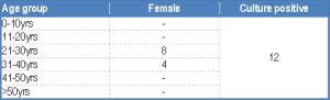
Total 30 cases were studied for superficial fungal infection of nail, culture positivity was found in 16 cases. Most common isolates were Candida albicans followed by Aspergillus fumigatus and Trichophyton rubrum. Males were most commonly affected than females. Candida albicans was isolated in cases of proximal subungal onychomycosis, Trichophyton rubrum was isolated in case of distal and lateral subungal onychomycosis and Aspergillus fumigatus was isolated in cases of white superficial onychomycosis. Right thumb was the commonest finger nail involved and among the toenail right big toe nail involvement was the commonest. Veer & Patwardhan (2005) from Aurangabad (Maharashtra) showed, most common isolates were dermatophytes from non-dermatophyte moulds and Candida albicans. Among dermatophytes, T. rubrum was the commonest etiological agent followed by T. mentagrophytes. Amongst the non-dermatophyte moulds, Aspergillus spp. was the most prevalent species. Kaur & Kashyap (2000-2005) from New Delhi showed, T. mentagrophytes and T. rubrum was the most commonly isolated dermatophytes followed by Aspergillus spp. and Candida albicans [29-31] . Sujatha & Grover (2000) from Bengaluru (Karnataka) observed most common isolated pathogen was Aspergillus niger, followed by T. rubrum, E. floccosum and C. albicans. The difference in the isolation pattern of fungi from the superficial fungl infection cases can be explained to some extent on the basis of climatic difference and relative humidity. Total 22 clinically suspected cases of otomycosis were studied. Culture positivity was found in 15 cases. Most common isolates were Aspergillus fumigates followed by Aspergillus niger. Males and females were equally affected. Following studies correlated with the present study: Chander & Maini (1996) from Chandigarh (Hariana) showed in their study that male-female ratio was equal. Most common isolated fungi were Aspergillus niger, followed by A. flavus and A. fumigatus. Kaur & Mittal (2000) from New Delhi observed, most predominant isolated fungi were A. fumigatus and A. niger [32-34] . 14 clinically suspected cases of vaginal candidiasis and 4 clinically suspected cases of oral candidiasis were studied. Candida albicans was isolated in 12 cases of vaginal candidacies and 2 cases of oral candidacies respectively. Vaginal candidacies were mainly found in pregnancy. Clinical and Laboratory standard Institute (CLSI) has recommended methodologies to be used for yeasts and filamentous fungi susceptibility testing. These methodologies gave reproducible results. E-test, Colorimetric testing, Flow cytometry have emerged as alternative techniques to address issues in different laboratories. Studies from many laboratories have expressed their inability to determine anti-fungal susceptibility testing using CLSI methodology. This leads to dependence of such laboratories on reference laboratories for anti-fungal susceptibility results leading to delay and inappropriate treatment. RPMI is recommended by CLSI for anti-fungal susceptibility testing; however, it is a synthetic, expensive, not routinely used medium in clinical microbiology laboratories. Sabouraud’s dextrose broth is comparatively cheaper, generally available in all clinical microbiological laboratories. Sabouraud’s dextrose broth with 0.5% agar selected for semi-solid agar susceptibility testing, as they are well established fungal growth media. In addition, 100% correlation with CLSI with respect to sensitivity, susceptibility positive and negative predictive value [12-17,22-15] . SAAS method showed potential as a screen for anti-fungal susceptibility testing. Anti-fungal susceptibility testing of dermatophytes (Trichophyton rubrum) was done by semi-solid agar dilution method using ant-fungal drug terbinafine. All isolates were sensitive. MIC of all isolates were 0.25µg/ml. Study of Khan, Singhal & Mathur(2006) also showed similar results and correlated with the present study. Anti-fungal susceptibility testing of Aspergillus fumigatus and Aspergillus niger were done by semi-solid agar dilution method using anti-fungal drug itraconazole [35-38] . All isolates were sensitive [Table-11] . MIC of all isolates were 0.12μg/ml. Anti-fungal susceptibility testing of Candida albicans was done by disc-diffusion method using anti-fungal drug fluconazole (25µg). All isolates were sensitive [Table-12] . Diameter of zone of inhibition of all isolates were >19mm.The present study and observation correlated with the study of Pfaller, Diekema & Gibbs (2007) [10-14,31-38] .
Newer anti-fungal agents have increased the therapeutic options thereby leading to the demand for in vitro determination of anti-fungal susceptibility. But the application of in vitro anti-fungal susceptibility testing in clinical research and to help in guiding the anti-fungal therapy has been limited by a lack of reproducibility and uncertain clinical relevance. To reduce the time required for anti-fungal susceptibility testing of filamentous fungi, semi-solid agar susceptibility method is the best method. The test can be set up for all fungi including mould. No special expertise or expensive equipments are needed, because the procedure is simple and similar for all fungi.
[1] Kannan P., Janaki C., Selvi G.S. (2006) Indian Journal Medical Microbiology, 24(3), 212-15.
» CrossRef » Google Scholar » PubMed » DOAJ » CAS » Scopus
[2] Drouhet E. (2005) Medical Mycology Topley & Wilson’s Microbiology & Microbial Infections,10th edition, 5-10.
» CrossRef » Google Scholar » PubMed » DOAJ » CAS » Scopus
[3] Mandell G.L., Bennett J.E., Dolin R. (2005) Principles and Practice of Infectious Diseases, 6th edition, 2.
» CrossRef » Google Scholar » PubMed » DOAJ » CAS » Scopus
[4] Sobera J.O., Elewski B.E. 92003) Dermatology, I, 2nd Edition. 1174-76.
» CrossRef » Google Scholar » PubMed » DOAJ » CAS » Scopus
[5] Forbes B.A., Beena P.M. (2003) Indian Jr. of Dermatology, Venereology and leprology, 69(4), 281-83.
» CrossRef » Google Scholar » PubMed » DOAJ » CAS » Scopus
[6] Calderone R.A. (1989) Mycoses, 32(2), 12-17.
» CrossRef » Google Scholar » PubMed » DOAJ » CAS » Scopus
[7] Barbhuiya S.N., Das S.K., Ghosh A., Dey S.K., Lahiri A. (2002) Indian Jr. of Dermatology., 47(4), 221-23.
» CrossRef » Google Scholar » PubMed » DOAJ » CAS » Scopus
[8] Kumar K., Kindo A.J., Kalyani J., Anandam S. (2007) Jn. of Medicine, 1(2), 12-15.
» CrossRef » Google Scholar » PubMed » DOAJ » CAS » Scopus
[9] Jain N., Sharma M., Saxena V.N. (2008) Indian Jn. of Dermatology, Venerology and Leprology, 74(3), 274-75.
» CrossRef » Google Scholar » PubMed » DOAJ » CAS » Scopus
[10] Koneman E.W. (2006) Color Atlas and Text Book of Diagnostic Microbiology. 6th edition.,1002-1043.
» CrossRef » Google Scholar » PubMed » DOAJ » CAS » Scopus
[11] Milne L.J.R. (1996) Fungi. Mackie & MaCartney Practical Medical Microbiology. 14th edition, 695-712.
» CrossRef » Google Scholar » PubMed » DOAJ » CAS » Scopus
[12] Rex J.H., Pfaller M.A., Walsh T.J., Chaturvedi V., Ingroff A., Ghannoum M., et al. (2001) Clinical Microbiological Reviews, 1494, 643-58.
» CrossRef » Google Scholar » PubMed » DOAJ » CAS » Scopus
[13] Geramishoar M., Zomorodian K., Emami M., Tarazooei B., Saadat F. Iranian (2002) Jn. of Public Health, 31(3), 100-104.
» CrossRef » Google Scholar » PubMed » DOAJ » CAS » Scopus
[14] Rattan A. (1999) Indian Journal of Medical Microbiology, 17(3), 12-28.
» CrossRef » Google Scholar » PubMed » DOAJ » CAS » Scopus
[15] Khan S., Singhal S., Mathur T., Upadhyay D.J., Rattan A. (2006) Indian Jn. of Medical Microbiology, 24(3), 171-76.
» CrossRef » Google Scholar » PubMed » DOAJ » CAS » Scopus
[16] Lee S.C., Fung C.P., Lee N., See L., Huang J., Tsai C., et al. (2001) Journal of Clinical Microbiology, 39(4), 1615-17.
» CrossRef » Google Scholar » PubMed » DOAJ » CAS » Scopus
[17] Aktas E., Yigit N. (2009) Jn. of medical Mycology, 19(2), 122-125.
» CrossRef » Google Scholar » PubMed » DOAJ » CAS » Scopus
[18] Abanmi A., Bakheshwain S., El Khizzi N., Zouman Ar., Hantirah S., Al Harthif., et al. (2008) Indian Jn. of Dermatology, 47(30), 229-35.
» CrossRef » Google Scholar » PubMed » DOAJ » CAS » Scopus
[19] Araujo C.R., Miranda K.C., Fernandes O., Soares A.J., Silva M.(2009) Rev. Inst. Med. Trop. Sao Paolo, 51(1), 36-38.
» CrossRef » Google Scholar » PubMed » DOAJ » CAS » Scopus
[20] Mukherjee P.K., Leidich S.D., Isham N., Leitnar I., Ryder N.S., Ghannoum M. (2003) Antimicrobial Agents and Chemotherapy, 47(1), 82-86.
» CrossRef » Google Scholar » PubMed » DOAJ » CAS » Scopus
[21] Barry A.L., Brown S.D. (1996) Journal of Clinical Microbiology, 34(9), 2154-57.
» CrossRef » Google Scholar » PubMed » DOAJ » CAS » Scopus
[22] Pfaller M.A., Diekema D.J., Gibbs D.L., Newell V.A., Meis J.F., Gould I.M., et al. (2007) Jn. of Clinical Microbiology, 45(6), 1735-45.
» CrossRef » Google Scholar » PubMed » DOAJ » CAS » Scopus
[23] Hoffman and Pfaller (2001) Oharmacotherap, 21980, 2-6.
» CrossRef » Google Scholar » PubMed » DOAJ » CAS » Scopus
[24] Fasunla J., Ibekwe T., Onakoya P. (2007) Mycoses, 51(1), 67-70.
» CrossRef » Google Scholar » PubMed » DOAJ » CAS » Scopus
[25] Flores J.M., Astillo V.B., Franco F.C., Huata A.B. (2009) Journal of Infect. Dev. Ctries, 3(4), 313-17.
» CrossRef » Google Scholar » PubMed » DOAJ » CAS » Scopus
[26] Kaur R., Kashyap B., Bhalla P. (2008) Indian Jn. of Medical Microbiology, 26(2), 108-16.
» CrossRef » Google Scholar » PubMed » DOAJ » CAS » Scopus
[27] Mishra G.S., Mehta N., Pal M. (2004) Mycoses, 47(1), 82-84.
» CrossRef » Google Scholar » PubMed » DOAJ » CAS » Scopus
[28] Nelson M.N., Martin A.G., Heffermasl M.P., Dermatology. 6th edition, II, Mc112.
» CrossRef » Google Scholar » PubMed » DOAJ » CAS » Scopus
[29] Veer P., Patwardhan N.S., Damle A.S. (2007) Indian Journal of medical Microbiology, 25(1), 53-56.
» CrossRef » Google Scholar » PubMed » DOAJ » CAS » Scopus
[30] Jesudanam T.M., Rama Rao G.R., Joga Lakshmi D., Ratna Kumari G. (2002) Indian Jn. of Dermatology, Venereology and Leprology, 68, 326-29.
» CrossRef » Google Scholar » PubMed » DOAJ » CAS » Scopus
[31] Sujatha V., Grover S., Dash K., Singh G. (2000) Indian Jn. of Dermatology, Venereology and Leprology, 66(5), 238-40.
» CrossRef » Google Scholar » PubMed » DOAJ » CAS » Scopus
[32] Kaur R., Kashyap B., Bhalla P.A. (2007) Indian Jn. of Dermatology, 52(1), 39-42.
» CrossRef » Google Scholar » PubMed » DOAJ » CAS » Scopus
[33] Garg A., Venkatesh V., Singh M., Pathak K.P., Kaushal G.P. (2004) Indian Jn. of Dermatology, 43(7), 498-502.
» CrossRef » Google Scholar » PubMed » DOAJ » CAS » Scopus
[34] Gupta M., Sharma N.L., Kanga A.K., Mahajan V.K., Tegta G.R. (2007) Indian Jn. of Dermatology, Venereology and Leprology, 73(6), 389-92.
» CrossRef » Google Scholar » PubMed » DOAJ » CAS » Scopus
[35] Chander J., Maini S., Subrahmanyan S., Handa A. (1996) Mycopathologia, 135(1), 9-12.
» CrossRef » Google Scholar » PubMed » DOAJ » CAS » Scopus
[36] Kaur R., Mittal N., Kakkar M., Aggarwal A.K., Mathur M.D. (2000) Ear, Nose and Throat Journal, 79(8), 606-9.
» CrossRef » Google Scholar » PubMed » DOAJ » CAS » Scopus
[37] Kemna M.E., Elewski B.E. (1996) Journal of American Acad. Dermatol., 35(4), 539-42.
» CrossRef » Google Scholar » PubMed » DOAJ » CAS » Scopus
[38] Rogers D., Kilkenny M., Marks R. (1996) Austales Journal of Demerol, 37(4), 178-84.
» CrossRef » Google Scholar » PubMed » DOAJ » CAS » Scopus
 | Table 1- Age-wise distribution pattern |
 | Table 2- Sex-wise distribution pattern |
 | Table 3- Results of KOH preparation |
 | Table 4- Results of culture study |
 | Table 5- Correlation between KOH and Culture study in the sample of skin scrapings |
 | Table 6- Age wise distribution of different clinical types in the sample of hair clipping |
 | Table 7- Sex wise distribution of different clinical types in the sample of hair clipping |
 | Table 8- Results of KOH preparation and culture study in the sample of hair clipping |
 | Table 9- Age distribution pattern of culture positive cases |
 | Table 10- Anti-fungal susceptibility testing of dermatophytes(Trichophyton rubrum) by semi-solid agar dilution method using anti fungal drug terbinafine |
 | Table 11- Anti-fungal susceptibility testing of Aspergillus fumigatus by semi-solid agar dilution method using anti-fungal drug Itraconazole |
 | Table 12- Anti-fungal susceptibility testing of Candida albicans by Disc diffusion method using anti-fungal drug Fluconazole (25µg) |