ISSN : 0975-5276
EISSN : 0975-9174

HEMASHENPAGAM N.1*
1Department of Microbiology, Hindushtan College of Arts and Science, Coimbatore-28, India
* Corresponding Author : drhemashenpagam@gmail.com
Received : 21-11-2011 Accepted : 09-12-2011 Published : 12-12-2011
Volume : 3 Issue : 3 Pages : 148 - 156
Int J Microbiol Res 3.3 (2011):148-156
DOI : http://dx.doi.org/10.9735/0975-5276.3.3.148-156
A total of 25 different types of actinomycetes were isolated from the soils of Agricultural University. All the isolated actinomycetes were characterized and identified based on the morphological, biochemical, cultural characteristics .The isolates were belonged to Streptomyces, Micromonospora and Nocardia species. Optimization for the growth of all the isolates was carried out in batch culture. Strain was cultured on the basal medium with different carbon and nitrogen sources and their effect on the growth was studied. Both primary and secondary screening methods were used to screen actinomycetes for antibacterial activity. The result of the screening revealed that all the isolates were against bacterial culture. But the best strain was found to be Streptomyces sp as they showed broad spectrum activity with big zone of inhibition, after mutation, the strain Streptomyces sp showed increased antibacterial activity against all the tested human bacterial pathogens. The crude extract were further analysed by thin layer chromatography on silica plates. The Rf values in the TLC chromatograph further characterize that Streptomyces sp 2comes under aminoglycoside group, very much related to Streptomycin and the Streptomyces sp 3 comes under the antibiotic cephalexin. HPLC profile of the antimicrobial compounds of Streptomyces sp were performed by Rheodysne Column (C-18) up to 10 min at 220 nm. The peaks which had antimicrobial activity of Streptomyces sp1 was identified to be at 3.296. This is similar to oxohexaene antibiotic whereas, the antimicrobial compound of Streptomyces sp3 showed a retention time of 3.233 on HPLC, this peak was similar to cephalexin.
Actinomycetes, mutation, TLC, HPLC.
Actinomycetes are gram positive bacteria frequently filamentous and sporulating with DNA rich in G+C from 55-75% [1] . The name actinomycetes derived from Greek aktis (a ray) and mykes (fungus) was given to these organisms from initial observation of their morphology. Streptomyces is the dominant among actinomycetes. They are responsible for the production of about half of the discovered bioactive secondary metabolites [2] , antibiotics [3] , antitumor agents [4] , immunosuppressive [5] and enzymes [6] . The diversity of terrestrial Actinomycetes are of extraordinary significance in several areas of science and medicine, particularly in antibiotic production [7] .
Actinomycetes are unparalleled sources of bio-active metabolites including antibiotics, plant growth factors, and other substances [8] . Streptomyces and other actinomycetes are major contributors to biological buffering of soils and have roles in organic matter decomposition conductive to crop production [9] . Actinomycetes are known to produce bioactive substances, especially antibiotics that are effective against phytopathogenic fungi [10] .
The present study is aimed to isolate, identify and to optimize the nutritional and cultural characterization of actinomycetes obtained from soil sample and then to compare the effect of mutation on antimicrobial activity of actinomycetes with mutated and non mutated strains. Finally to purify antibacterial metabolites from efficient strain and to detect antimicrobial compounds by HPLC.
The rhizosphere soil samples were collected from 10 different localities of Agricultural University. 25 different soil samples were collected from the surface to about 10 cm deep as a mixture for the isolation of actinomycetes.
Actinomycetes were isolated by spread plate technique following the serial dilution of soil samples on starch casein agar [11] plates containing cycloheximide and nystatin (each at concentration of 50 µg/ml of medium) and incubated at 28°C for 7 days. The following media were also used for the isolation of actinomycetes: Starch caesin agar, Starch caesin nitrate agar, Actinomycetes Isolation agar, Oat meal agar etc.
Isolation plates were incubated at 28-30˚C for 7-15 days for fast growing actinomycetes or up to 35 days for slow growing ones. Identification of Actinomycetes to genus level was conducted by first using morphological and chemical criteria according to Bergey’s Manual of Determinative Bacteriology [12] .
50 ml of the basal medium was distributed into each of 250 ml Erlenmeyer flask and sterilized. The carbon and nitrogen sources were sterilized separately and added just prior to inoculation. Starch, Glucose, Fructose, Maltose, Mannitol were added separately as carbon sources into the basal medium at 1% concentration. Various nitrogen sources such as casein, yeast extract, peptone, soya bean meal, NH4Cl and NH4 NO3 were provided separately into the basal medium at 1% concentration. Five ml of spore suspension of strain was inoculated into the carbon and nitrogen supplemented basal medium separately and incubated at 28°C for seven days at 220 rpm in a rotary shaker. The respective biomass production was recorded.
A modified cross- streak method [13] was used for antimicrobial activity .Single streak of actinomycetes was made on surface of the modified nutrient agar and incubated at 28°C. [14] After observing a good ribbon like growth of the actinomycetes on the plates, the overnight pathogenic bacterial strains, such as Escherichia coli, Staphylococcus aureus, Salmonella typhi, Shigella sp, Klebsiella pneumonia, Proteus vulgaris which were isolated from the admitted patients (PSG Hospital) were streaked at right angles to the original streak of actinomycetes and incubated at 28°C and the incubation distance was measured after 24-48 hrs. A control plate was also maintained without inoculating the actinomycetes, to assess the normal growth of bacteria.
The filtrate was subjected for solvent extraction method to recover antibacterial metabolites in pure form [15] . Ethyl acetate was added to the filtrate in the ratio of 1:1(v/v) and shaken vigorously for 1 h for complete extraction. The ethyl acetate phase contains antibiotic substances separated from aqueous phase. It was evaporated to dryness in water bath and the residue obtained was used to determine antimicrobial activity [16] .
The antimicrobial activity was determined by agar well method [17] purified extract obtained by the evaporation of the ethyl acetate extract. Then 25, 50, 75 and 100µl of it was loaded into well bored and test organism (0.5 McFarland turbidity standard) were swabbed on Muller Hinton agar plates. The plates were incubated at 37°C for 18-24 hours and examined. The diameter of the zones of complete inhibition was measured to the nearest whole millimeter. The formation of inhibition zone around the pathogenic strains is due to the production of secondary metabolites by actinomycetes isolates.
The strains which showed efficient antimicrobial activity were further selected to study the effect of mutation on their antibiotic production. The selected strains were cultured in the tubes containing 9 ml Yeast Extract Malt Broth. The tubes were inoculated with one loopful of the strain and incubated in a rotatory shaker at 30°C for 96 hours. After incubation, the tubes were removed from the shaker and 3 ml of each culture was exposed to UV irradiation at a distance of 30 cm for 180 sec. One ml of the exposed cultures was transferred to 9 ml of Glycerol starch Broth medium and the tubes were incubated for 96 hours on a shaker at 30°C. After incubation, the tubes were removed from the shaker and the broth was centrifuged at 2000 rpm for 20 min and the supernatant was used to examine the post mutation effect on the strains for their antibacterial activity [18] .
Optimization of mobile phase (butanol: acetic acid: water in two ratios of 4:1:5 and 2:1:8) for known antibiotics and test antibiotics was done by using 7.6 x 2.4 cm silica gel plates, prepared and activated at 110 ˚C for half an hour. Chromatogram was developed by loading 10 μl each fraction and running for half an hour. Spots on the plates were visualized in an iodine chamber [19,20] .
The culture filtrate was extracted three times by the same volume of ethyl acetate. After the removal of cell mass, supernatant was analysed by spectrophotometer at 220nm. This was carried out to for HPLC analysis. Culture filtrate extracted by ethyl acetate was analyzed by HPLC. 10 µl of sample was injected to C18 column (250mm X 4.6mm X 5mm). The flow rate was 0.50 ml/min. Sample was analysed at 220 nm wavelength.
In order to purify the active fraction (methanol: chloroform 20: 80 extract); chloroform was added gradually (dropwise) until formation of the first precipitate. The precipitate was separated by centrifugation. Each fraction was tested in inhibition test. Ammonium sulphate was also used for the extraction. Ammonium sulfate was grind in a glass mortar to be a fine powder that easily to dissolve.
This powder was added gradually to the antagonistic solution. The solution was shacked using a vortex. The formed precipitate was separated by centrifugation. The amount of ammonium sulfate was noted for each precipitate and calculated as saturation percent. Each precipitate was dissolved in water and re-precipitated again by ammonium sulfate as a purification step [21] . Each precipitate was dissolved in methanol and centrifuged to remove any residue of ammonium sulfate. This step was repeated several times until the removing all amounts of ammonium sulfate. Each precipitate was used for the inhibition test and analysed by HPLC.
The present study was undertaken in 10 different localities of Agricultural University. 25 different soil samples were collected from surface of about 10 cm deep as a mixture for the isolation of actinomycetes [Fig-1] .
The cultural characterization of the actinomycetes isolates were studied by using different types of solid media. The result indicated that all the tested media supported the growth and significant result was produced in Starch casein nitrate agar. [Table 1] , [Fig-1] .
The results on morphological, physiological and biochemical activity revealed that the isolated strains of actinomycetes belonged to Streptomyces sp, Micromonospora sp and Nocardia sp. [Table-2] [Plate-1] , [Plate-2] .
Biochemical test were performed like Indole, MR,VP,Citrate, Nitrate reduction, Urease, Catalase, Oxidase, Starch Hydrolysis, Gelatin Hydrolysis, Lipid Hydrolysis, Caesin Hydrolysis, TSI and the result were tabulated. [Table-3] [22] .
Based on the above biochemical characteristics the strains were identified as Micromonospora sp, Streptomyces sp, Nocardia sp.
Utilization of carbon sources namely Starch, Glucose, Fructose, Maltose, Mannitolwere tested for all the strains [Table-4] .
All the actinomycete strains produced highest biomass, when glucose was supplemented in the medium.
Utilization of nitrogen sources namely Casein, Yeast extract, Peptone, Soya bean meal, NH4Cl, NH4 NO3 were tested for all the strains [Table-5] .
All the actinomycetes isolates showed antibacterial activity against pathogenic E.coli, Salmonalla typhii, Shigella, Klebsiella pneumonia, Proteus vulgaris, Staphylococcus aureus in primary screening and all the species were found to be more resistant. The control plate was done without the actinomycetes [Plate-3] .
Out of the six screened strain, Streptomyces sp showed a very broad spectrum of antimicrobial activity against both gram positive, gram negative and fungal pathogens used in the test. The antagonistic activity of all the actinomycetes isolates were shown in the [Table 6] .
The strains which showed efficient antibacterial activity were further selected to study the effect of mutation on their antibiotic production [Table-7] , [Fig-1] .
The mutated strains were checked for their antibacterial activity against human pathogens namely E.coli, Staphylococcus, Salmonella, Shigella, Klebsiella, Bacillus subtilis. But only Streptomyces sp 1 and Streptomyces sp 2 showed increase in inhibition zone against E.coli, Pseudomonas, Staphylococcus, and Streptococcus.
When visualized under iodine vapour each extract showed two spots. One at the point of loading itself and other near to the solvent front on the chromatogram. The retention factor of the moved spot is given in the [Table-8] .
HPLC is being routinely used for the analytical estimation of various antibiotics. In the present investigation, HPLC profile of the antimicrobial compounds of Streptomyces sp were performed by Rheodysne Column (C-18) up to 10 min at 220 nm.
The antimicrobial compounds of Streptomyces sp1 showed absorption peaks at retention time (min) 3.296, 3.740, 3.856, 4.444, 5.112, 5.340 and 6.406. Similarly, the antimicrobial compounds of Streptomyces sp 2 showed absorption peaks at retention time (min) 2.964, 3.233, 4.102, 4.272, 5.080, 5.301. [Fig-2] , [Fig-3] , [Table-9] , [Table-10] .
The peaks which had antimicrobial activity of Streptomyces sp1 was identified to be at 3.296. This is similar to oxohexaene antibiotic [23] .
Whereas, the antimicrobial compound of Streptomyces sp3 showed a retention time of 3.233 on HPLC, this peak was similar to cephalaxine [24] .
25 different types of actinomycetes were isolated from the soils of Agricultural University. Isolation of actinomycetes has always been faced with difficulties in comparison to their competitors like other bacteria and fungi [25] . This may be due to their long incubation period. Use of selective media (Starch casein media) incorporated with antibiotics, cycloheximide (50μg/ ml) and nystatin (50μg/ ml) was crucial inhibiting contaminating microorganisms. The cultural characterization of the actinomycetes isolates were studied by using different types of solid media. Here the growth of actinomycetes is found to be abundant in Starch caesin nitrate agar medium [Table-1] .
The result on biochemical characterization indicated that pigment production was very well observed in all the strains. Based on the morphological, Physiological and biochemical characteristics, the purified isolates of actinomycetes belonged to Streptomyces sp. As they showed good sporulation with compact, chalk-like dry colonies of different colony variation from pink to white color [Table-2] . All the isolates were found to be gram positive organism and they showed a branched mycelium in their cell morphology similar to fungal characters. Most of the isolates were efficient in hydrolysing starch and casein [26] except a few strains. Indole production was strictly negative but catalase was positive in all the isolates. Production of hydrogen sulphide, gelatin hydrolysis, caesin and starch hydrolysis showed a positive result in majority of the isolates [Table-3] .
Optimization for the growth of all the isolates was carried out in batch culture. Strain was cultured on the basal medium with different carbon sources and their effect on the growth was studied. The strain was able to grow in all the tested carbon sources. All the actinomycete strains produced highest biomass, when glucose was supplemented in the medium. Addition of other carbon sources such as starch, fructose, maltose, Mannitol, etc. to the medium also favoured the growth but the growth was less when compared with glucose [Table-4] . All the actinomycetes strain produced highest biomass, when peptone was used as nitrogen source and among inorganic nitrogen sources ammonium chloride showed moderate effect on the growth of the isolate [Table-5] . It is clear from the results that the growth was greatly influenced by the nature and type of the nitrogen source supplied in the culture medium [27] .
Both primary and secondary screening methods were used to screen actinomycetes for antibacterial activity. The first screening was used to select the antibacterial isolates and determine the range of microorganism that were sensitive to the antibiotic. The secondary screening method was crucial to select the isolates for further studies. The result of the screening revealed that all the isolates were against bacterial culture. But the best strain was found to be Streptomyces sp as they showed broad spectrum activity with big zone of inhibition. Therefore the isolates were chosen for fermentation. The antibacterial metabolites from fermented broth were extracted in organic solvent (ethyl acetate) by solvent extraction method [28] .
In this study after mutation, the strain Streptomyces sp showed increased antibacterial activity against all the tested human bacterial pathogens. The mutation is responsible for the increased production of antibiotics. Zone of inhibition was observed against E.coli (2.5 cm), Pseudomonas (1.8 cm), Staphylococcus (2.2 cm), Streptococcus (1.8 cm). [18] .
The crude extract were further analysed by thin layer chromatography on silica plates using butanol: acetic acid: water in two ratios of 4:1:5 as solvent system and standard antibiotics were loaded. Each extract produced two spots when the chromatogram was visualized under iodine vapour. One spot at the sample loading and the other was near the solvent front with Rfvalue of 1.00 for Streptomyces sp1, 0.06 for Streptomyces sp2, 0.53 for Streptomyces sp3, 1.09 for Nocardia sp1, 0.26 for Nocardia sp2, 1.00 for Micromonospora sp. The test compound was relatively compared with the known antibiotics. The Rf values in the TLC chromatograph further characterize that Streptomyces sp 2 comes under aminoglycoside group, very much related to Streptomycin and the Streptomyces sp 3 comes under the antibiotic cephalexin [29] .
HPLC is being routinely used for the analytical estimation of various antibiotics. In the present investigation, HPLC profile of the antimicrobial compounds of Streptomyces sp wereperformed by Rheodysne Column (C-18) up to 10 min at 220 nm. The antimicrobial compounds of Streptomyces sp1 showed absorption peaks at retention time (min) 3.296, 3.740, 3.856, 4.444, 5.112, 5.340 and 6.406. Similarly, the antimicrobial compounds of Streptomyces sp 2 showed absorption peaks at retention time (min) 2.964, 3.233, 4.102, 4.272, 5.080, 5.301. The peaks which had antimicrobial activity of Streptomyces sp1 was identified to be at 3.296. This is similar to oxohexaene antibiotic [23] Whereas, the antimicrobial compound of Streptomyces sp3 showed a retention time of 3.233 on HPLC, this peak was similar to cephalexin, a semisynthetic derivative of cephalosporin C [24] produced by S. clavuligenus [30] .
The present study was an attempt to identify and pick out versatile actinomycetes strains that display antimicrobial activity against a variety of microbial pathogens intrinsically.
Our result indicate that we are able to identify two different antibiotics namely cephalaxine, oxohexaene. Most microorganisms have developed resistance to existing antibiotics. So it has provoked the need of constant research like ours on the production of newer antibiotics to overcome the resistant microorganism.
Further investigation is needed in order to determine the structure of active compounds and to scale up the production.
[1] Ho C., Lo C., Lai N., Cheah H. and Wong N. (2002) ASEAN Rev. Biodiversity Environ. Conser. 9:1-7.
» CrossRef » Google Scholar » PubMed » DOAJ » CAS » Scopus
[2] Berdy J. (2005) Review article. J Antibiot, 58: 1–26.
» CrossRef » Google Scholar » PubMed » DOAJ » CAS » Scopus
[3] Strohl W.R. (2004) Antimicrobials. In Microbial Diversity and Bioprospecting. Edited by Bull AT. ASM Press; 336-355.
» CrossRef » Google Scholar » PubMed » DOAJ » CAS » Scopus
[4] Cragg G.M., Kingston D.G.I., Newman D.J. (2005) (Eds). Anticancer Agents from Natural Products. Taylor & Francis, 23:676-686.
» CrossRef » Google Scholar » PubMed » DOAJ » CAS » Scopus
[5] Mann J. (2001) Nat Prod Rep, 18:417-430
» CrossRef » Google Scholar » PubMed » DOAJ » CAS » Scopus
[6] Oldfield C., Wood N.T., Gilbert S.C, Murray F.D., Faure F.R. (1998) Antonie Van Leeuwenhoek, 74:119-132.
» CrossRef » Google Scholar » PubMed » DOAJ » CAS » Scopus
[7] Magarvey N.A., Keller J.M., Berman V., Dworkin M. and Sherman D.H. (2004) Applied and environmental microbiology, 70(12): 7520-7529.
» CrossRef » Google Scholar » PubMed » DOAJ » CAS » Scopus
[8] Shahidi B.G.H., Fooladi M.H., Mahdavi M.J., Shahghasi A. (2004) Biotechnol. 3:126-130.
» CrossRef » Google Scholar » PubMed » DOAJ » CAS » Scopus
[9] Dhingra O.D., Sinclair J.B., (1995) Basic Plant Pathology Methods. CRC Press: USA. 287-296, 390-391.
» CrossRef » Google Scholar » PubMed » DOAJ » CAS » Scopus
[10] Crawford D.L., Lynch J.M., Whipps J.M., Ousley M.A. (1993) Appl. Environ. Microbiol. 59: 3899-3905.
» CrossRef » Google Scholar » PubMed » DOAJ » CAS » Scopus
[11] Williams S.T. and F.L. Davies (1965) Journal of General Microbiology 38: 251-261.
» CrossRef » Google Scholar » PubMed » DOAJ » CAS » Scopus
[12] Holt J.G., Krieg N.R., Sneath P.H.A., Stanley J.T. and Williams S.T. (1994) Bergey’s manual of Determinative Bacteriology, 9th edn, pp 571-701, Williams & Wilkins, Baltimore.
» CrossRef » Google Scholar » PubMed » DOAJ » CAS » Scopus
[13] Lakshmanaperumalsamy P. (1978) Studies on Actinomycetes with special reference to antagonistic Streptomyces from sediments of Porto Novo coastal zone. Doctoral thesis, Annamalai Univ. India.
» CrossRef » Google Scholar » PubMed » DOAJ » CAS » Scopus
[14] Balagurunathan R. (1992) Antagonistic Actinomycetes from Indian shallow sea sediments with reference to a alpha, beta unsaturated gamma lactose type of antibiotic from Streptomyces griseobrunneus Doctoral thesis, Annamalai Univ. India.
» CrossRef » Google Scholar » PubMed » DOAJ » CAS » Scopus
[15] Liu C.M., Westley J.W., Herman T.E., Prasser B.L.T., Palleroni N., Evans R.H. and Miller P.A. (1986) Journal of Antibiotics, 39 (12): 1712-1718.
» CrossRef » Google Scholar » PubMed » DOAJ » CAS » Scopus
[16] Westley J.W., Evans R.H., Sello L.H., Troupe N., Liu C.M. and Blount J.F. (1979) J. Antibiot 32:100-107.
» CrossRef » Google Scholar » PubMed » DOAJ » CAS » Scopus
[17] Parekh J. and Chanda S.V. (2007) Turk. J. Biol. 31: 53-58.
» CrossRef » Google Scholar » PubMed » DOAJ » CAS » Scopus
[18] Philips J.N. (1960) J. Genetics, 46:317-322.
» CrossRef » Google Scholar » PubMed » DOAJ » CAS » Scopus
[19] Busti E., Monciardini P., Cavaletti L., Bamonte R., Lazzarini A. and Sosio (2006) Microbiology, 152:675-683.
» CrossRef » Google Scholar » PubMed » DOAJ » CAS » Scopus
[20] Thangadurai S., Shukla S.K. and Anjaneyulu Y. (2002) Analytical Science, 18:97-100.
» CrossRef » Google Scholar » PubMed » DOAJ » CAS » Scopus
[21] Shawn Doonan (1991) Protein purification protocols. Humana Press, Totowa, New Jersey.
» CrossRef » Google Scholar » PubMed » DOAJ » CAS » Scopus
[22] Holt J.G. (1989) Bergey's manual of systematic bacteriology, Williams S.T. and Sharpe M.E., Baltimore, Md: Williams and Williams.
» CrossRef » Google Scholar » PubMed » DOAJ » CAS » Scopus
[23] Harindran J., Gupte T.E., Naik S.R. (1999) World J. Microbiol Biotechnol 15: 425-430.
» CrossRef » Google Scholar » PubMed » DOAJ » CAS » Scopus
[24] Aharonowitz Y., Demain A.L. (1978) Antimicrob agents chemother, 14: 159-164.
» CrossRef » Google Scholar » PubMed » DOAJ » CAS » Scopus
[25] Williams S.T. and Cross T. (1971) Actinomycetes In: Methods in Microbiology, Booth C. (eds). Academic press, London.
» CrossRef » Google Scholar » PubMed » DOAJ » CAS » Scopus
[26] Ravel J., Wellington W.H. and Hill R.T. (2000) Applied Environ. Microbiol, 66:529-534.
» CrossRef » Google Scholar » PubMed » DOAJ » CAS » Scopus
[27] Yu J., Liu Q., Liu Q., Liu X., Sun Q., Yan J. (2008) BioresTechnol, 99: 2087-91.
» CrossRef » Google Scholar » PubMed » DOAJ » CAS » Scopus
[28] Manjula, Rajaguru C.P. and Muthuselvam M. (2009) Adv. Biol. Res, 3:84-88.
» CrossRef » Google Scholar » PubMed » DOAJ » CAS » Scopus
[29] Brooks G.H., Butel J.S. and Morse S.A. (2001) Antimicrobial chemotherapy. In: Medical microbiology (22nd edition) (eds. Jawetz, Melnick & Adelberg). International edition. Lange Medical books/McGraw Hill Publication.
» CrossRef » Google Scholar » PubMed » DOAJ » CAS » Scopus
[30] Nakagawa T., Haginaka J., Yamaoka K. and Uno T. (1978) J. Antibiot; 31: 769-775.
» CrossRef » Google Scholar » PubMed » DOAJ » CAS » Scopus
 | Fig. 1- Percentage of actinomycetes on different localities of Agricultural university, Coimbatore |
 | Fig. 1a- Effect of mutation |
 | Fig. 2- Chromatogram of antimicrobial compound of Streptomyces sp1 |
 | Fig. 3- Chromatogram of antimicrobial compound of Streptomyces sp3 |
 | Plate 1- Gram staining of Streptomyces sp1, sp2 and sp3 |
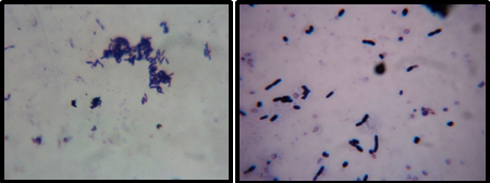 | Plate 2- Gram staining of Nocardia sp1, 2 |
 | Plate 3- Primary Screening of actinomycetes |
 | Table 1- Cultural characterization of actinomycetes +++ Abundant; ++ Good; + Moderate |
 | Table 2- Morphological Characterization of actionmycetes |
 | Table 3- Biochemical characterization of actinomycetes + Positive, - Negative |
 | Table 4- Carbon source utilization for selected isolates Excellent ++++, Good +++, Fair ++, Poor +, Nil - |
 | Table 5- Nitrogen source utilization of selected isolates Excellent ++++, Good +++, Fair ++, Poor +, Nil |
 | Table 6- Antagonistic activity of Actinomycetes isolates against human pathogen in 100 μl of extract |
 | Table 7- Effect of mutation on antibacterial activity |
 | Table 8- Results for TLC Rfvalues of all the strains were recorded and they are compared with the standard antibiotics |
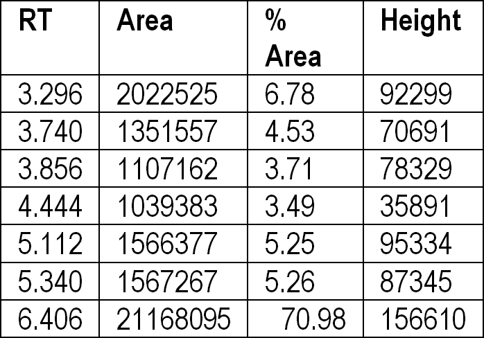 | Table 9- Chromatogram values of Streptomyces sp 1 |
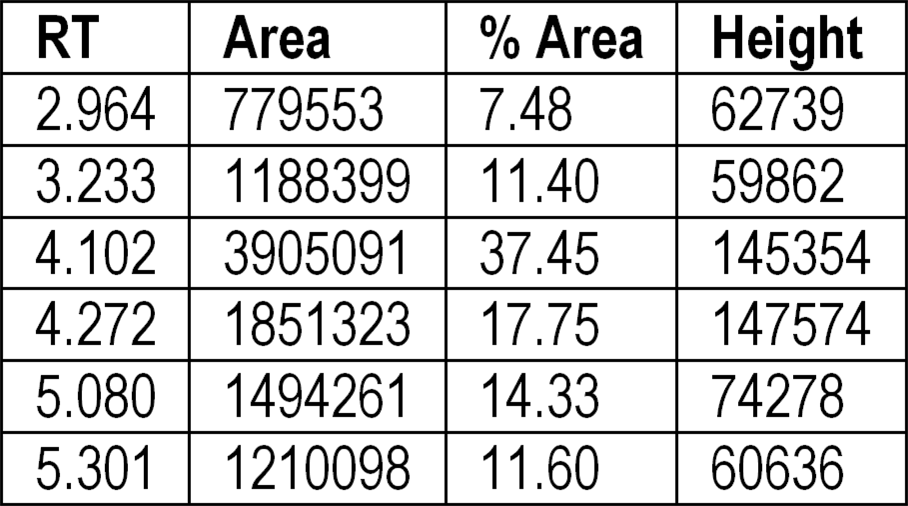 | Table 10- Chromatogram of antimicrobial compound of Streptomyces sp3 |