ISSN : 0976-5530
EISSN : 0976-5549

DEEPA PARVATHI V.1*, PRIYANKA V2
1Department of Human Genetics, Sri Ramachandra University, Porur, Chennai 600116
2Department of Human Genetics, Sri Ramachandra University, Porur, Chennai 600116
* Corresponding Author : deepa_305@yahoo.com
Received : 25-08-2011 Accepted : 27-09-2011 Published : 29-09-2011
Volume : 2 Issue : 1 Pages : 23 - 28
Int J Med Clin Res 2.1 (2011):23-28
DOI : http://dx.doi.org/10.9735/0976-5530.2.1.23-28
Conflict of Interest : None declared
Tumor cells have an altered amino acid metabolism. They express high levels of tryptophan and L-arginine metabolizing enzymes. This leads to depletion of tryptophan and L-arginine and locally blocks T cell proliferation. Therefore, the characteristic energy metabolism of tumor cells leads to immune suppression and contributes to immune escape processes at the tumor site [1]. Excess dietary Tryptophan has shown to cause increased tumor incidence in tumorous strains of Drosophila [2]. Canton (wild type) flies were exposed to 10mM, 20mM, 30mM, 40mM and 50mM concentrations of Tryptophan. Phenotypic analysis of the exposed flies and qualitative analysis of the isolated DNA was done 24 hours and 48 hours after exposure to Tryptophan. The quality of the DNA was evaluated using Nanodrop and the DNA was subjected to Fragmentation assay to study the damage induced. Phenotypic changes observed were elongated abdomen with distinct curling and discoloration of thorax to mild orange. The isolated DNA was found to be of good quality and the fragmentation assay showed patterns of shearing. As the damage observed was not significant and the tumor induction was not observed in the experimental concentrations, tryptophan was considered non tumorigenic at the above said concentrations. However, higher concentrations of Tryptophan may induce tumor.
Tryptophan, Tumor induction, Drosophila, DNA Damage.
Drosophila melanogaster, has been used in research in genetics and is a common model organism in developmental biology. The entire genus contains about 1,500 species which are diverse in appearance, behavior and breeding habitat [3] Drosophila melanogaster is amenable to genetic analysis because of the ease with which it can be grown and the size of its genome, as they have about 13,000 genes on 4 chromosomes.
They have short generation time and a high fecundity rate. They have easily observed body plan development in embryonic, larval, and adult stages which can be easily, identified using a light microscope. Changes in these features reflect mutations in genes controlling differentiation and developmental processes [4-5] .
Cancer involves the cooperation between mutations in several genes, and unique combinations of signaling pathways can be coerced for novel outputs. In vivo, specific pathways are involved in tumorigenesis in different tissues, and provide evidence that the interactions between the tumor and its microenvironment are highly important in cancer development. Simpler animal model systems that mimic some of the steps of mammalian tumorigenesis can provide a more rapid means to uncover novel pathways of cancer. One such model organism that has been widely studied is Drosophila melanogaster [6] . Tryptophan serves as the precursor for the synthesis of Serotonin (5-hydroxy tryptamine / 5-HT) and Melatonin (N-acetyl-5-methoxytryptamine). Serotonin is synthesized through a 2-step process involving the enzyme Tryptophan hydroxylase and a decarboxylation catalyzed by Decarboxylase [7] . [Fig-1] .
As a consequence of malignant transformation tumor cells show an altered metabolic phenotype. A link between tumor metabolism and cancer was first described many years ago by Warburg (aerobic glycolysis, "Warburg effectâ€) and this "glycolytic phenotype†seems to be necessary for the evolution of invasive human cancers. In addition, tumor cells have an altered amino acid metabolism. They express high levels of tryptophan and L-arginine metabolizing enzymes. This leads to depletion of tryptophan and L-arginine and locally blocks T cell proliferation. Therefore the characteristic energy metabolism of tumor cells leads to immune suppression and contributes to immune escape processes at the tumor site [8] .
Tryptophan is metabolized through serotonin, indole, and kynurenine (KN) pathways. Uptake of an excess amount of tryptophan may result in the accumulation of higher concentrations of metabolites mainly from the KN pathways in the bladder. These metabolites could interact with nitrite to become mutagenic nitrosamines. They could be a promoter in the initiator–promoter model of carcinogenesis. They produced bladder cancer when implanted in the bladder. They also interact with transition metals copper or iron to form reactive radicals or reactive oxygen species (ROS) [9] . High free tryptophan (F-TRP) plasma levels are found in cancer patients. F-TRP plasma concentrations are affected by the levels of its carrier, albumin (ALB), and free fatty acids (FFA) competing with TRP for ALB binding sites. The lack of correlation between F-TRP, ALB and FFA in cancer patients has shown a tumor-dependent effect on the rise in F-TRP [10-11] .
1.02115g of L-Tryptophan dissolved in 100ml of distilled water.
10mM - 200μl of 0.5M stock solution
20mM - 400μl of 0.5M stock solution
30mM - 600μl of 0.5M stock solution
40mM - 800μl of 0.5M stock solution
50mM - 1000μl of 0.5M stock solution
• Tris HCL - 100mM
• EDTA - 100mM
• NaCl - 100mM
• SDS - 0.5%
Canton (wild type) flies were allowed to breed in corn meal agar until the desired number of flies have obtained.
Different concentrations of Tryptophan (10mM, 20mM, 30mM, 40mM and 50mM) were each mixed with 3g of instant food. Control and Test were set up for each concentration. Two vials of instant food (one for 24 hours exposure and one for 48 hours exposure) were prepared for each concentration.
The food was labeled with appropriate concentrations and the duration of exposure. It was then foiled and allowed to set for 2 hours at 25 °C (to avoid fermentation). About 30 flies were exposed to each concentration of Tryptophan and observed under the microscope (for phenotypic changes) after 24 hours and 48 hours of exposure.
The exposed flies were transferred to a culture vial and placed in a beaker containing ice in the refrigerator for 30 minutes. After the flies have collapsed, they were transferred into a mortor and pestle and crushed by adding 500µl of Solution A.
The contents were then transferred into an eppendorf and incubated in a water bath set to 70 °C for 30 minutes. DNA was then isolated using PCI method.
The quality of DNA was checked using Nanodrop Spectrophotometer. About 1.5µl of the isolated DNA samples were added to the pedestals of the Nanodrop and the results were analyzed.
The quality checked DNA samples were subjected to Fragmentation assay, to study the damage, by subjecting the samples to 1.5% Agarose gel electrophoresis for 45 minutes and the results were documented.
[Table-1] & [Table-2] Exposure to tryptophan:
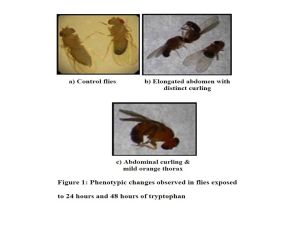
Male and female flies were exposed to varying concentrations of Tryptophan (10mM, 20mM, 30mM, 40mM, and 50mM) and the phenotypic changes were observed microscopically. [Fig-2]
[Table-3] & [Table-4] DNA isolation:
DNA was isolated from both Control and Exposed flies using Phenol Chloroform method and the quality of the DNA was checked using Nanodrop. The Nanodrop results show that the DNA was found to be of good quality.
DNA from Control and Tryptophan exposed flies were subjected to Fragmentation assay to study the damage. The results obtained were documented [Fig-2] & [Fig-3] . Distinct shearing was observed in all concentrations (10mM, 20mM, 30mM, 40mM and 50mM) of Tryptophan after 24 hours exposure. Whereas, a single band (at 200bp) was observed in 10mM, 20mM and 30mM concentrations and mild shearing was observed in 40mM and 50mM concentrations. [Fig-3] , [Fig-4] .
The present study indicates that Tryptophan is capable of inducing minimum amount of damage to the DNA at the given concentrations. Canton flies (both males and females) were exposed to varying concentrations of Tryptophan and analyzed for phenotypic changes and the quality of the DNA obtained from the exposed flies was checked. Uptake of excess dietary Tryptophan has reported to increase tumor incidence in tumor strains of Drosophila. Although Canton strains exposed to different concentrations of Tryptophan did not show tumor formation, few abnormalities such as discoloration of thorax, elongation of abdomen and curling of abdomen were observed in both 24 hours as well as 48 hours exposed flies. Flies exposed to 50mM concentration of Tryptophan, showed 80% and 50% viability after 24 hours and 48 hours of exposure respectively. Further, the flies analyzed under the stereo zoom microscope for phenotypic changes revealed elongation of abdomen, curling of abdomen and discoloration of thorax. The difference in viability might be due to the prolongation of exposure to Tryptophan which proves the lethality of Tryptophan at such high concentrations. The quality of DNA obtained from exposed flies was found to be of good quality using Nanodrop, thus proving no phenol contamination and the yield of DNA being high. Fragmentation assay of DNA obtained from flies exposed to Tryptophan for 24 hours showed distinct shearing at all the observed concentrations. Significant shearing was observed in 20mM, 30mM, 40mM and 50mM concentrations of Tryptophan after 24 hours exposure whereas, at 10mM concentration, mild shearing was observed. At concentrations of 10mM, 20mM and 30mM, a single band was observed and at 40mM and 50mM concentrations mild shearing was observed after 48 hours of exposure. As only a single band was observed, it cannot be indicated as a fragment. As no significant damage has been observed, shearing cannot be employed as a measure of the damage caused to the DNA. Hence, advanced methods including protein profiling by SDS PAGE and Wing Spot Assay may be required to assess the total damage caused to the DNA upon exposure. Also, higher concentrations of Tryptophan (>50mM) should be analyzed for DNA damage and tumorigenic activity.
[1] Kreutz M., Andreesen R. (1990) Blood, 76, 2457-2461.
» CrossRef » Google Scholar » PubMed » DOAJ » CAS » Scopus
[2] Burnet B., Sang J.H. (1968) Genetics, 211-235.
» CrossRef » Google Scholar » PubMed » DOAJ » CAS » Scopus
[3] Rubin G.M., Lewis E.B. (2000) Science, 287 (5461), 2216-2218.
» CrossRef » Google Scholar » PubMed » DOAJ » CAS » Scopus
[4] Adams M.D., Celniker S.E., Holt R.A., Evans C.A., Gocayne J.D. (2000) Science, 287 (5461), 2185-2195.
» CrossRef » Google Scholar » PubMed » DOAJ » CAS » Scopus
[5] Klug W.S., Cummings M.R., Spencer C.A., Palladino M.A. Concepts of genetics, Pearson Benjamin Cummings.
» CrossRef » Google Scholar » PubMed » DOAJ » CAS » Scopus
[6] Brumby A.M., Richardson H.E. (2005) Nature, 5 (8), 629-639.
» CrossRef » Google Scholar » PubMed » DOAJ » CAS » Scopus
[7] Turner E.H., Blackwell A.D. (2005) Medical Hypotheses 65 (1), 138–144.
» CrossRef » Google Scholar » PubMed » DOAJ » CAS » Scopus
[8] Rabinowitz J.D., White E. (2010) Science 330 (6009), 1344-1348.
» CrossRef » Google Scholar » PubMed » DOAJ » CAS » Scopus
[9] Chung K.T., Gadupudi G.S. (2011) Environmental Molecular Mutagen, 52 (2), 81-104.
» CrossRef » Google Scholar » PubMed » DOAJ » CAS » Scopus
[10] Cascino A., Cangiano C., Ceci F., Franchi F., Mineo T., Mulieri M., Muscaritoli M., Fanelli R.F. (1991) Anticancer Research, 11 (3), 1313-1316.
» CrossRef » Google Scholar » PubMed » DOAJ » CAS » Scopus
[11] Burnet B., Sang J.H. (1964) Genetics, 223-235.
» CrossRef » Google Scholar » PubMed » DOAJ » CAS » Scopus
 | Fig. 1- |
 | Fig. 2- |
 | Fig. 3- |
 | Fig. 4- |
 | Table 1- Exposure to tryptophan – 24 hours |
 | Table 2- Exposure to tryptophan – 48 hours |
 | Table 3- Nanodrop results for 24 hours exposure to tryptophan |
 | Table 4- Nanodrop results for 48 hours exposure to tryptophan |